
















Copyright: ©Author(s) 2026.
World J Gastrointest Surg. Jun 27, 2026; 18(6): 118552
Published online Jun 27, 2026. doi: 10.4240/wjgs.118552
Published online Jun 27, 2026. doi: 10.4240/wjgs.118552
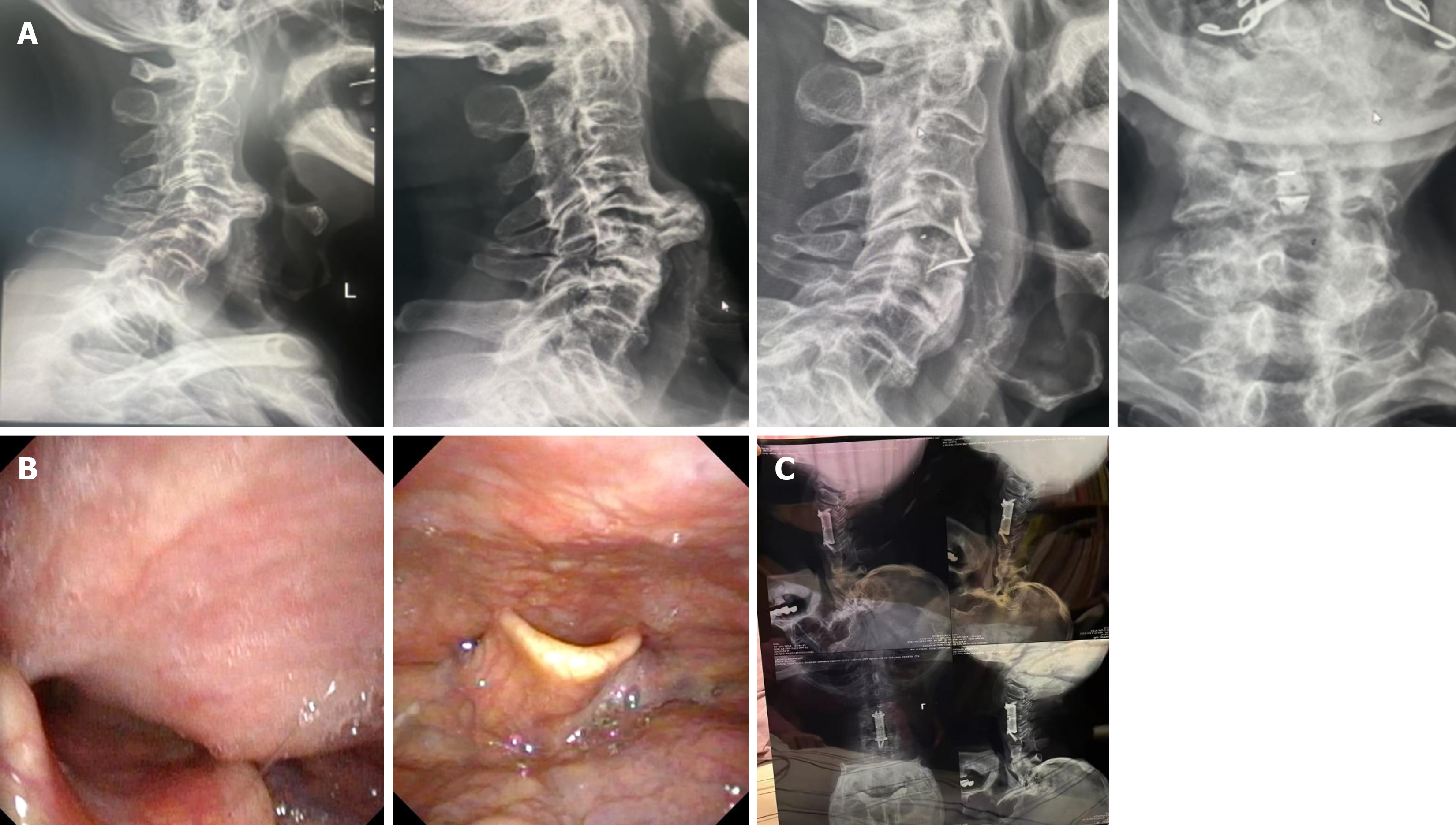
Figure 3 Imaging examinations.
A: Case 1: Postoperative imaging examinations indicated complete removal of the hyperplastic bone at the anterior edge of the C3/4 vertebral bodies; B: Case 1: Nasopharyngoscopy before (left) and after (right) surgery illustrated the resolution of the extrinsic compression/bulge on the posterior pharyngeal wall; C: Case 2: Postoperative imaging performed after 1 year revealed the resolution of the cervical and thoracic vertebral osteophytes.
- Citation: Li XH, Sun YY, Chen TT, Yang F, Lai ZB, He J. Surgical management of dysphagia at the oropharyngeal-esophageal junction due to cervical spondylosis: Two case reports. World J Gastrointest Surg 2026; 18(6): 118552
- URL: https://www.wjgnet.com/1948-9366/full/v18/i6/118552.htm
- DOI: https://dx.doi.org/10.4240/wjgs.118552