
















Copyright: ©Author(s) 2026.
World J Gastrointest Surg. Jun 27, 2026; 18(6): 117909
Published online Jun 27, 2026. doi: 10.4240/wjgs.117909
Published online Jun 27, 2026. doi: 10.4240/wjgs.117909
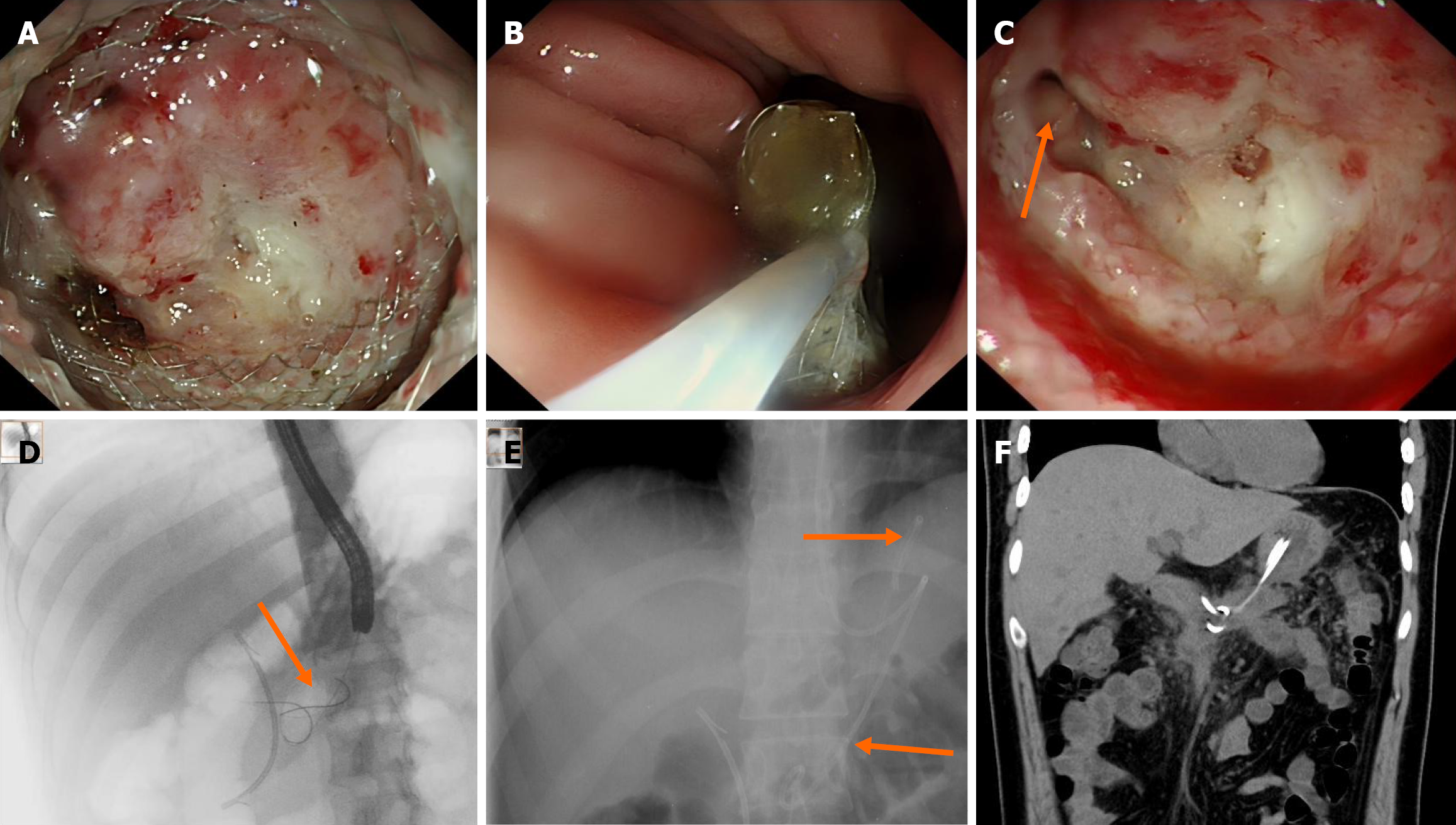
Figure 5 Management of branch cavity ostium occlusion by lumen-apposing metal stents edge with plastic stents placement.
A: Endoscopic view showing sealed lumen-apposing metal stents cavity-side opening; B: Lumen-apposing metal stents removal using a snare; C: Small fistula with purulent discharge at the 9 o’clock position of the cavity wall; D: Fluoroscopic confirmation of guidewires access into recurrent collection (arrow indicates guidewires); E: Fluoroscopic view showing nasobiliary drainage tube and double-pigtail stent placement (arrows indicate devices); F: Follow-up computed tomography demonstrating proper stents position and near-complete resolution of the collection.
- Citation: Liao DX, Yang L, Gou HX, Li X, Xie Q, Luo H, Wen Y. Successful redrainage after failure of lumen-apposing metal stents by addressing flange-induced ostial occlusion: A case report. World J Gastrointest Surg 2026; 18(6): 117909
- URL: https://www.wjgnet.com/1948-9366/full/v18/i6/117909.htm
- DOI: https://dx.doi.org/10.4240/wjgs.117909