
















Copyright: ©Author(s) 2026.
World J Gastrointest Surg. Jun 27, 2026; 18(6): 117526
Published online Jun 27, 2026. doi: 10.4240/wjgs.117526
Published online Jun 27, 2026. doi: 10.4240/wjgs.117526
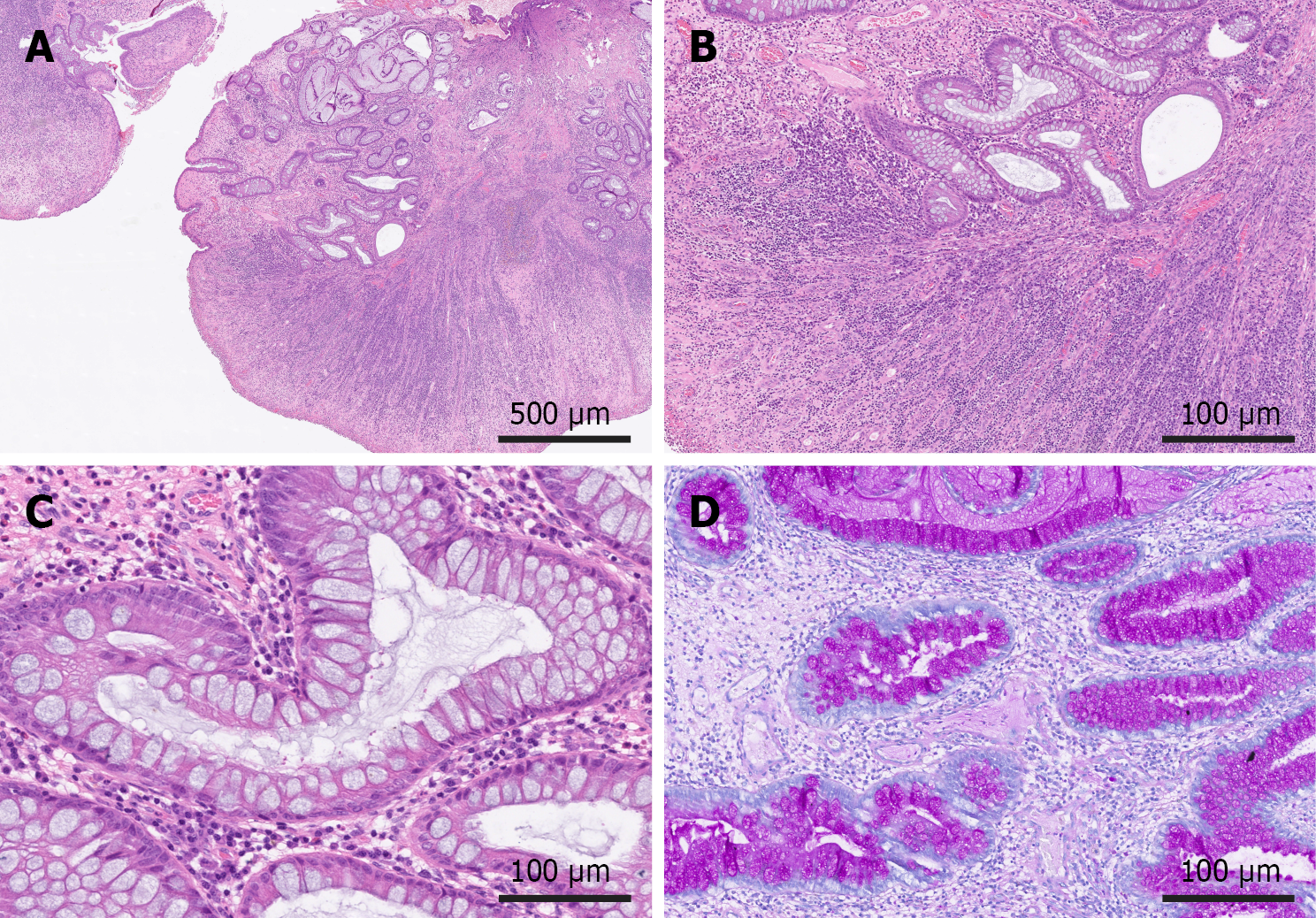
Figure 2 Characteristic histopathological features of cap polyposis.
A-C: Hematoxylin and eosin staining. Erosive mucosal surface covered by thick inflammatory granulation tissue (pseudomembrane) with architectural disarray and prominent cystic dilation of crypts (A). Dense mixed inflammatory cell infiltration within the lamina propria (B). Elongated, dilated, and distorted crypts with a preserved glandular architecture and an absence of significant cytological atypia (C); D: Periodic acid-Schiff reaction. Periodic acid-Schiff reaction highlighting the fibrin-rich pseudomembrane overlying the mucosa.
- Citation: Shan LN, Shan JL, Yin SJ, Bai BJ, Xu K, Dai S, Zhou W. Clinical features, diagnosis, and treatment of colorectal cap polyposis. World J Gastrointest Surg 2026; 18(6): 117526
- URL: https://www.wjgnet.com/1948-9366/full/v18/i6/117526.htm
- DOI: https://dx.doi.org/10.4240/wjgs.117526