
















Copyright: ©Author(s) 2026.
World J Gastrointest Surg. May 27, 2026; 18(5): 119504
Published online May 27, 2026. doi: 10.4240/wjgs.v18.i5.119504
Published online May 27, 2026. doi: 10.4240/wjgs.v18.i5.119504
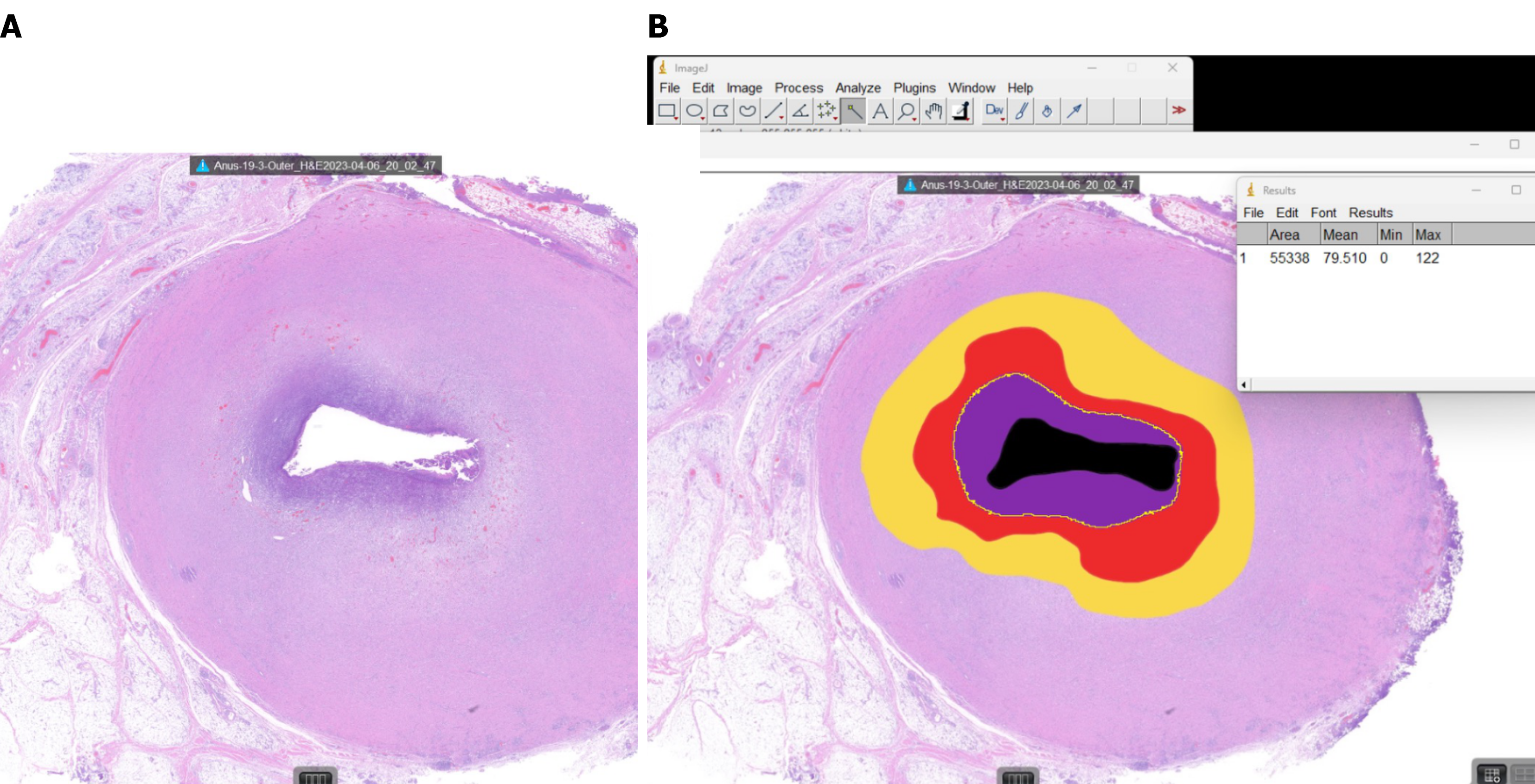
Figure 3 Representative histological section illustrating Image J-based quantification of ablation and granulation tissue.
A: Hematoxylin and eosin -stained cross-section of an outer fistula-tract segment harvested after radiofrequency ablation ablation (120 °C/24 seconds), showing a centrally obliterated lumen surrounded by a concentric zone of thermal injury; B: The same section processed in Image J, with color overlays delineating tissue compartments used for morphometry: Residual lumen (black), carbonized/charred core (violet), coagulative necrosis (red), transition zone (orange) and residual granulation tissue (yellow).
- Citation: Yoon S, Ji WB, Um JW, Hong KD. Optimizing radiofrequency ablation parameters for perianal fistula treatment: A porcine model study. World J Gastrointest Surg 2026; 18(5): 119504
- URL: https://www.wjgnet.com/1948-9366/full/v18/i5/119504.htm
- DOI: https://dx.doi.org/10.4240/wjgs.v18.i5.119504