
















Copyright: ©Author(s) 2026.
World J Gastrointest Surg. May 27, 2026; 18(5): 119504
Published online May 27, 2026. doi: 10.4240/wjgs.v18.i5.119504
Published online May 27, 2026. doi: 10.4240/wjgs.v18.i5.119504
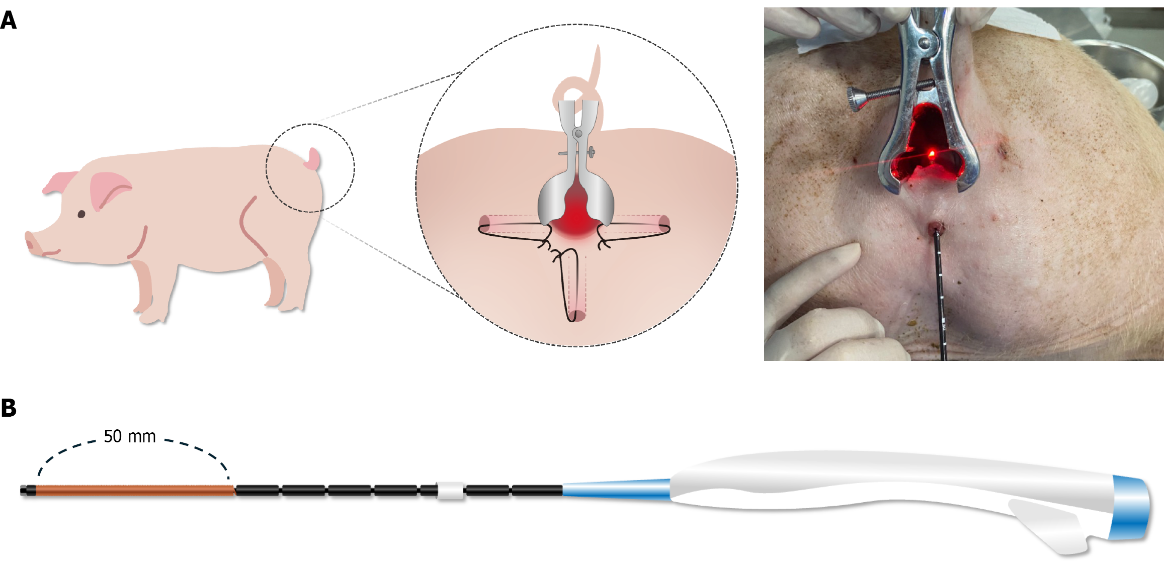
Figure 1 Experimental set-up and novel radiofrequency ablation device.
A: Schematic of the porcine fistula model used for in-vivo optimization of radiofrequency ablation (RFA) parameters. Three fistula tracts were surgically created in each animal and kept patent with setons, as illustrated in the enlarged inset. For ablation, seton was removed and the RFA catheter introduced endoluminally, as confirmed by the intra-operative photograph; B: Prototype segmental RFA catheter. The distal 50 mm copper-colored segment constitutes the active ablation blade, and graduations at 10 mm intervals along the shaft allow precise depth control. This 50 mm active segment delivers a uniformly distributed ablation along its entire length, thereby overcoming the focal-tip limitation of conventional devices.
- Citation: Yoon S, Ji WB, Um JW, Hong KD. Optimizing radiofrequency ablation parameters for perianal fistula treatment: A porcine model study. World J Gastrointest Surg 2026; 18(5): 119504
- URL: https://www.wjgnet.com/1948-9366/full/v18/i5/119504.htm
- DOI: https://dx.doi.org/10.4240/wjgs.v18.i5.119504