
















Copyright: ©Author(s) 2026.
World J Gastrointest Surg. May 27, 2026; 18(5): 118166
Published online May 27, 2026. doi: 10.4240/wjgs.v18.i5.118166
Published online May 27, 2026. doi: 10.4240/wjgs.v18.i5.118166
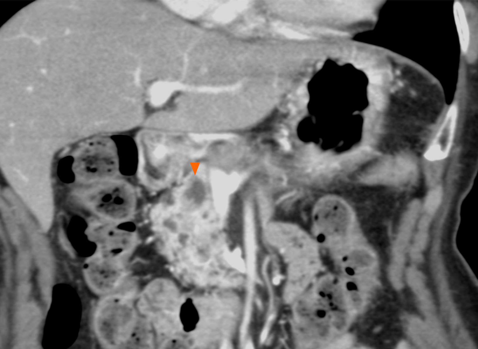
Figure 3 Imaging findings before the second surgery.
Contrast-enhanced computed tomography image obtained in June 2007 before the second surgery. The main pancreatic duct in the remnant pancreatic head is dilated (orange arrowhead), leading to the diagnosis of main duct-type intraductal papillary mucinous neoplasm in the remnant pancreatic head.
- Citation: Nomoto K, Hishida M, Hayashi M, Nomoto S. Late nodal recurrence after two curative resections for microinvasive intraductal papillary mucinous carcinoma: A case report. World J Gastrointest Surg 2026; 18(5): 118166
- URL: https://www.wjgnet.com/1948-9366/full/v18/i5/118166.htm
- DOI: https://dx.doi.org/10.4240/wjgs.v18.i5.118166