
















Copyright: ©Author(s) 2026.
World J Gastrointest Surg. May 27, 2026; 18(5): 118166
Published online May 27, 2026. doi: 10.4240/wjgs.v18.i5.118166
Published online May 27, 2026. doi: 10.4240/wjgs.v18.i5.118166
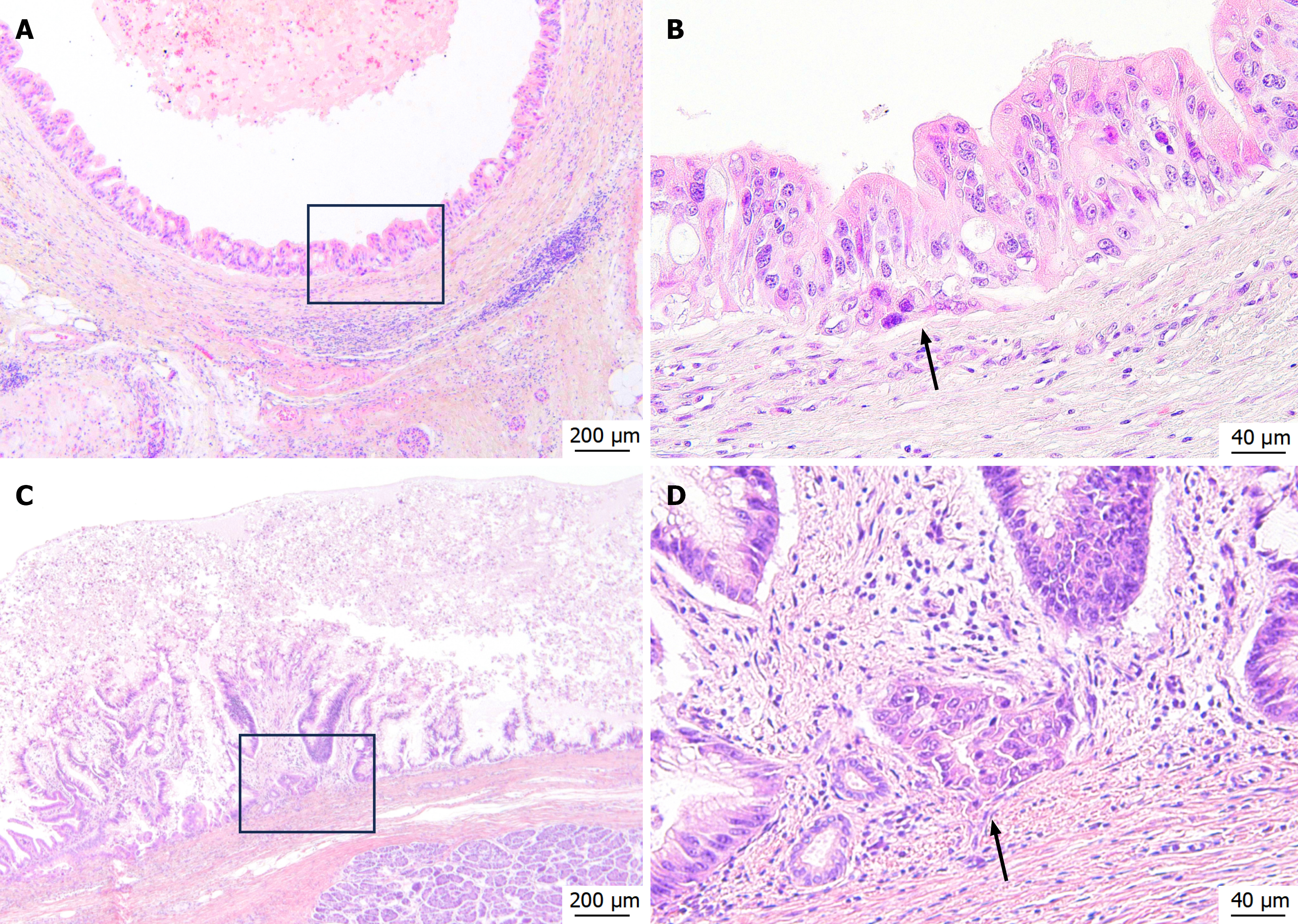
Figure 2 Histopathological findings of microinvasive intraductal papillary mucinous carcinoma (Hematoxylin and eosin staining).
A and B: Histopathological images from the first surgery. Atypical columnar epithelium showing dysplasia proliferates in papillary patterns within the dilated pancreatic duct. Slight irregularity of the basal aspect indicating microinvasion is observed in the marginal area (black arrow) (A: × 40; B: × 200); C and D: Histopathological images from the second surgery. Findings similar to those of the first surgery, including microinvasion (black arrow), are observed (C: × 40; D: × 200).
- Citation: Nomoto K, Hishida M, Hayashi M, Nomoto S. Late nodal recurrence after two curative resections for microinvasive intraductal papillary mucinous carcinoma: A case report. World J Gastrointest Surg 2026; 18(5): 118166
- URL: https://www.wjgnet.com/1948-9366/full/v18/i5/118166.htm
- DOI: https://dx.doi.org/10.4240/wjgs.v18.i5.118166