
















Copyright: ©Author(s) 2026.
World J Gastrointest Surg. May 27, 2026; 18(5): 117565
Published online May 27, 2026. doi: 10.4240/wjgs.v18.i5.117565
Published online May 27, 2026. doi: 10.4240/wjgs.v18.i5.117565
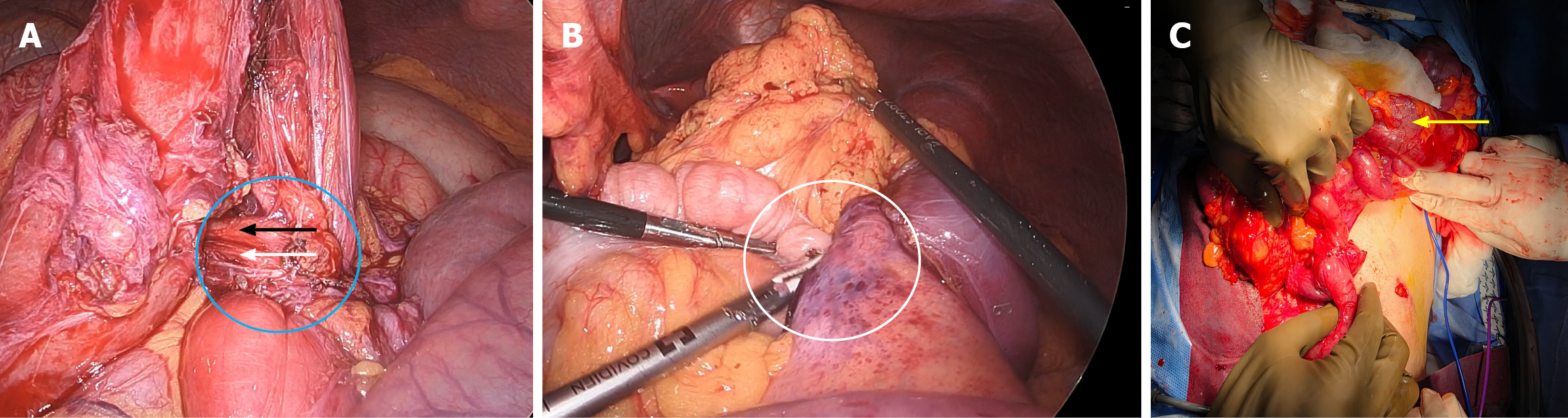
Figure 2 Intraoperative sequence demonstrating laparoscopic confirmation of aortomesenteric duodenal compression, identification of a concomitant right paraduodenal defect, and definitive reconstruction.
A: Laparoscopic view showing the third portion of the duodenum (blue circle) passing beneath the superior mesenteric artery (black arrow) and the superior mesenteric vein (white arrow), with extrinsic aortomesenteric compression causing focal luminal narrowing of third portion of the duodenum at the crossing; B: Laparoscopic view demonstrating the right paraduodenal (Waldeyer) defect, occupied by herniated transverse colon with adjacent bowel loops (white circle), such that the defect itself is not directly visualized because it is filled by the herniated intestinal contents; C: Open view following conversion to laparotomy, showing the completed side-to-side duodeno-duodenostomy (yellow arrow) restoring duodenal continuity.
- Citation: Sacasa F, Ploneda C, Cervantes M, Betancourt B, Casal J, Bedoya J, Valladares W. Superior mesenteric artery syndrome and right paraduodenal hernia causing intestinal obstruction during pregnancy: A case report. World J Gastrointest Surg 2026; 18(5): 117565
- URL: https://www.wjgnet.com/1948-9366/full/v18/i5/117565.htm
- DOI: https://dx.doi.org/10.4240/wjgs.v18.i5.117565