
















Copyright: ©Author(s) 2026.
World J Gastrointest Surg. May 27, 2026; 18(5): 117565
Published online May 27, 2026. doi: 10.4240/wjgs.v18.i5.117565
Published online May 27, 2026. doi: 10.4240/wjgs.v18.i5.117565
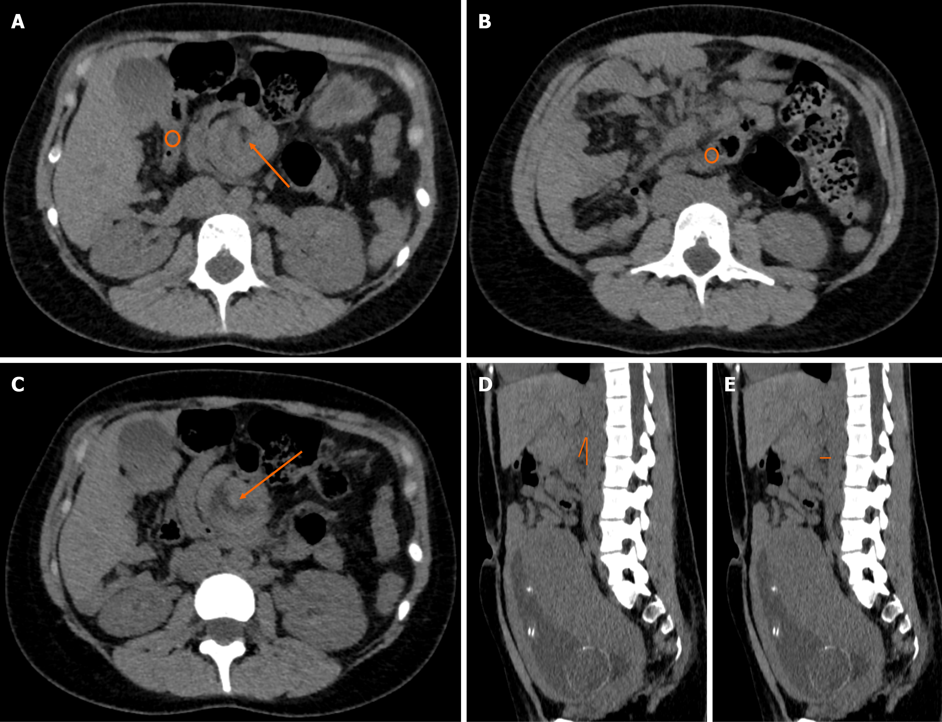
Figure 1 Non-contrast abdominal computed tomography in a 21-year-old primigravida demonstrating primary aortomesenteric (Wilkie’s) duodenal compression with a concomitant right paraduodenal hernia.
A-C: Axial images demonstrate a right paraduodenal hernia through Waldeyer’s fossa (orange arrow in A). The ascending colon and part of the hepatic flexure are contained within the hernia defect (orange circles in A and B). This anatomic distortion alters the course of the superior mesenteric artery, which is visualized centrally within the hernia defect (orange arrow in C). The third portion of the duodenum is poorly delineated and compressed at the aortomesenteric crossing, indicating that the duodenal obstruction arises from primary aortomesenteric compression rather than from the hernia itself; D and E: Sagittal reconstructions demonstrate a reduced aortomesenteric angle of approximately 19° (orange angle marker in D) and a reduced aortomesenteric distance of approximately 8 mm (orange arrow indicating the measurement in E). Because this pregnancy-adapted study lacked oral and intravenous contrast, these measurements are illustrative rather than diagnostic and are included to highlight the limitations of non-contrast computed tomography in pregnancy. Definitive confirmation of Wilkie’s syndrome was established intraoperatively.
- Citation: Sacasa F, Ploneda C, Cervantes M, Betancourt B, Casal J, Bedoya J, Valladares W. Superior mesenteric artery syndrome and right paraduodenal hernia causing intestinal obstruction during pregnancy: A case report. World J Gastrointest Surg 2026; 18(5): 117565
- URL: https://www.wjgnet.com/1948-9366/full/v18/i5/117565.htm
- DOI: https://dx.doi.org/10.4240/wjgs.v18.i5.117565