
















Copyright: ©Author(s) 2026.
World J Gastrointest Surg. May 27, 2026; 18(5): 115825
Published online May 27, 2026. doi: 10.4240/wjgs.v18.i5.115825
Published online May 27, 2026. doi: 10.4240/wjgs.v18.i5.115825
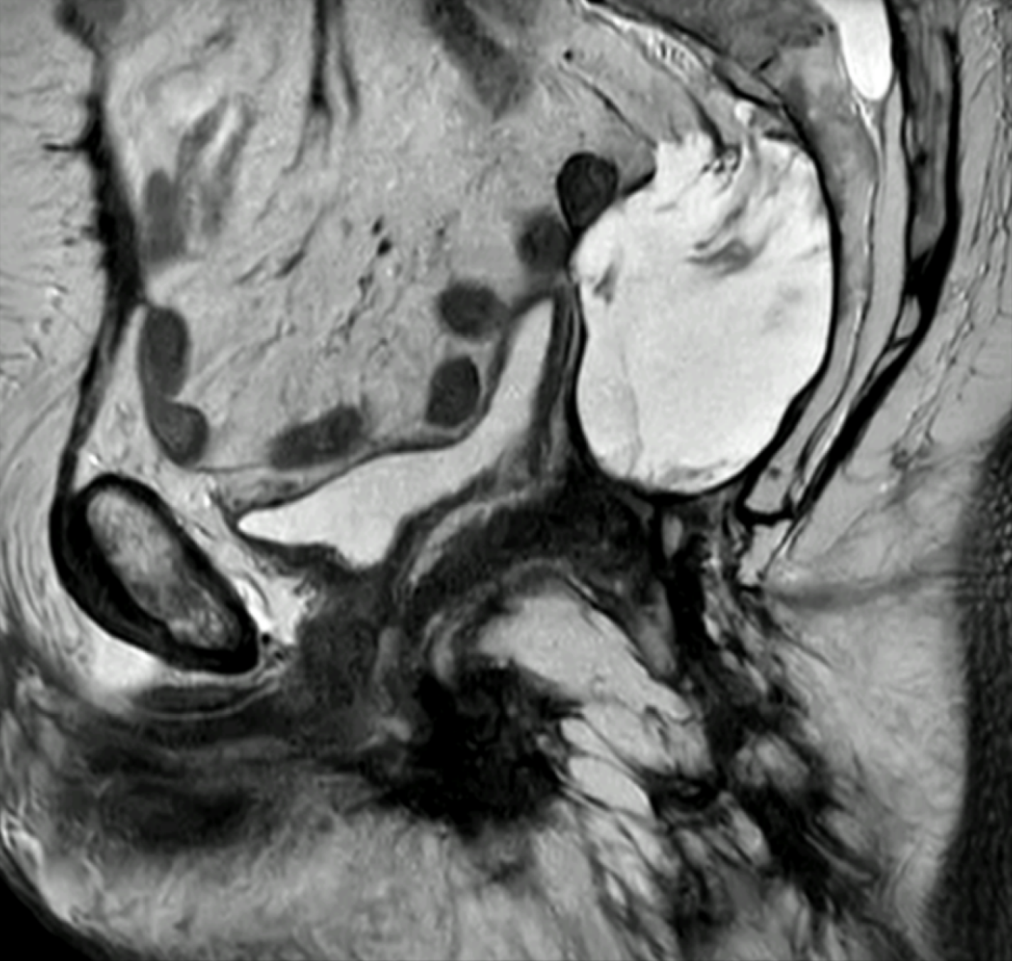
Figure 3 Pelvic magnetic resonance imaging revealed a local recurrence of mucinous adenocarcinoma 4 years subsequent to an R0 abdominoperineal resection.
Sagittal T2-weighted image demonstrated a lobulated, high-signal-intensity mass in the presacral space, consistent with recurrent mucinous disease. The lesion exhibited homogeneous T2 hyperintensity typical of extracellular mucin and ill-defined margins against adjacent pelvic structures. These imaging features are characteristic of mucinous local recurrence and help differentiate it from postoperative seroma or fibrosis.
- Citation: Patriti A, Graziano F, Chiari R. Mucinous rectal adenocarcinoma and neoadjuvant therapy: Implications for surgical practice. World J Gastrointest Surg 2026; 18(5): 115825
- URL: https://www.wjgnet.com/1948-9366/full/v18/i5/115825.htm
- DOI: https://dx.doi.org/10.4240/wjgs.v18.i5.115825