
















Copyright: ©Author(s) 2026.
World J Gastrointest Surg. Apr 27, 2026; 18(4): 116972
Published online Apr 27, 2026. doi: 10.4240/wjgs.v18.i4.116972
Published online Apr 27, 2026. doi: 10.4240/wjgs.v18.i4.116972
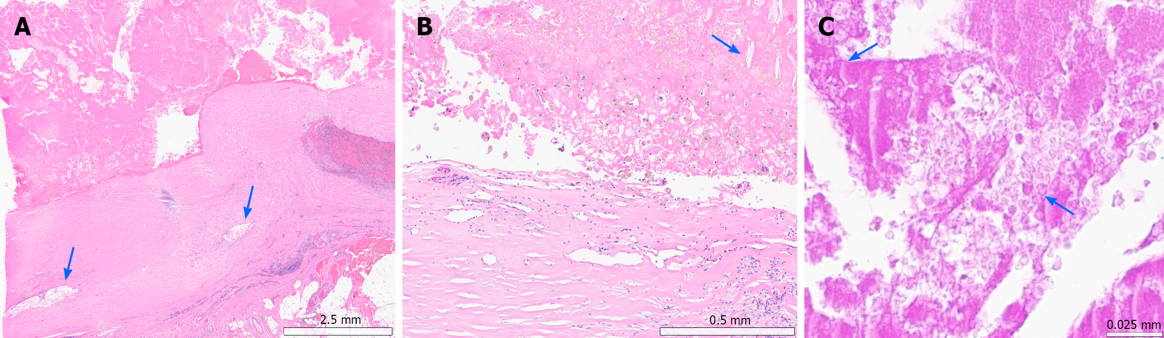
Figure 4 Histopathological features of the subserosal cystic lesion with foreign-body reaction A: Low-power microscopic examination demonstrates a thick fibrous cystic wall in the subserosa, filled with necrotic materials.
Cholesterol crystals (arrow) with an accompanying foreign-body reaction are noted; B: High-power magnification of the cyst wall reveals dense fibrosis with mild chronic inflammatory cell infiltration. The cystic cavity contains numerous macrophages, amorphous necrotic debris with cholesterol crystals (arrow); C: Periodic Acid-Schiff staining of the intracystic contents highlights irregularly shaped foreign materials (arrow).
- Citation: Song SH, Lee SS, Kim DJ. Transverse colon foreign body-associated reactive mass mimicking gastrointestinal stromal tumors: A case report. World J Gastrointest Surg 2026; 18(4): 116972
- URL: https://www.wjgnet.com/1948-9366/full/v18/i4/116972.htm
- DOI: https://dx.doi.org/10.4240/wjgs.v18.i4.116972