
















Copyright: ©Author(s) 2026.
World J Gastrointest Surg. Apr 27, 2026; 18(4): 115487
Published online Apr 27, 2026. doi: 10.4240/wjgs.v18.i4.115487
Published online Apr 27, 2026. doi: 10.4240/wjgs.v18.i4.115487
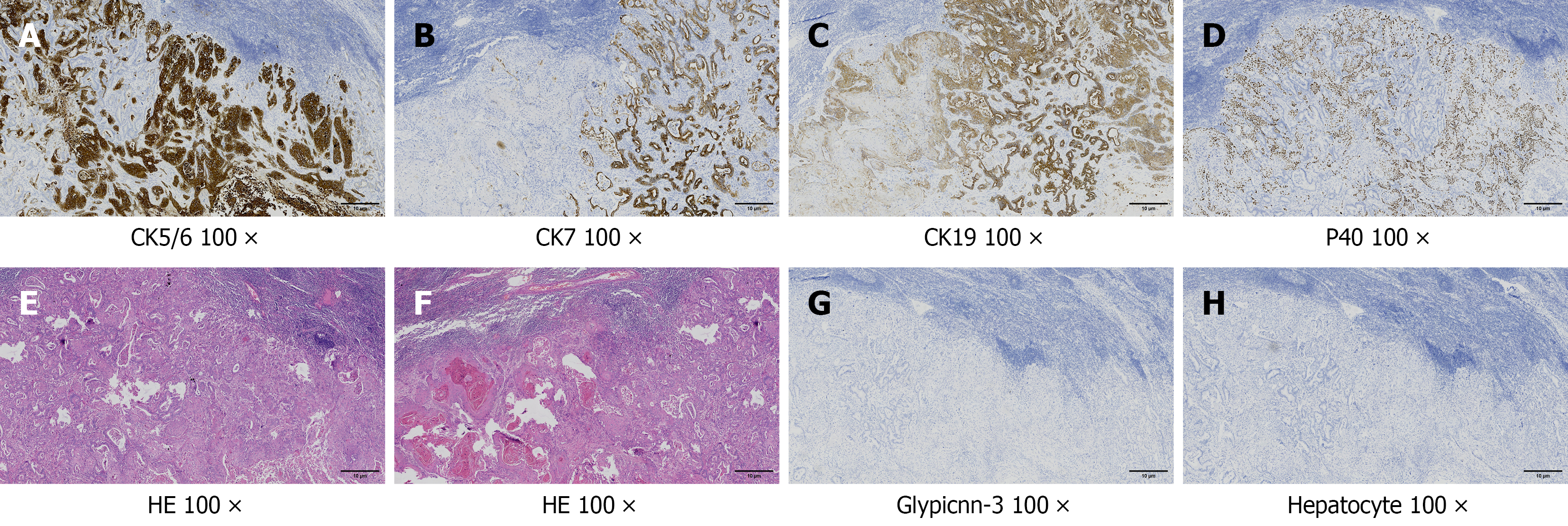
Figure 4 Pathological images of adenosquamous carcinoma in lymph node metastasis.
The adenocarcinoma component of the tumor has tubular structures, and the cells show a nest-like arrangement (squamous cell carcinoma component). A-C: Positive staining of cytokeratin (CK) 5/6, CK7 and CK19 shows brown and granular deposits in the cytoplasm (× 100); D: Positive staining of protein 40 shows brown and granular deposits in the nucleus (× 100); E and F: The tumor showed atypia, mitosis, solid nests, and tubular structures (hematoxylin and eosin stain, × 100); G and H: Negative staining of glypicnn-3 and hepatocytes in tumor cells (× 100). CK: Cytokeratin; P40: Protein 40; HE: Hematoxylin and eosin.
- Citation: Fu RB, Dong XF. Diagnostic challenges and multidisciplinary management of primary adenosquamous carcinoma of the liver: A case report and review of literature. World J Gastrointest Surg 2026; 18(4): 115487
- URL: https://www.wjgnet.com/1948-9366/full/v18/i4/115487.htm
- DOI: https://dx.doi.org/10.4240/wjgs.v18.i4.115487