
















Copyright: ©Author(s) 2026.
World J Gastrointest Surg. Apr 27, 2026; 18(4): 115487
Published online Apr 27, 2026. doi: 10.4240/wjgs.v18.i4.115487
Published online Apr 27, 2026. doi: 10.4240/wjgs.v18.i4.115487
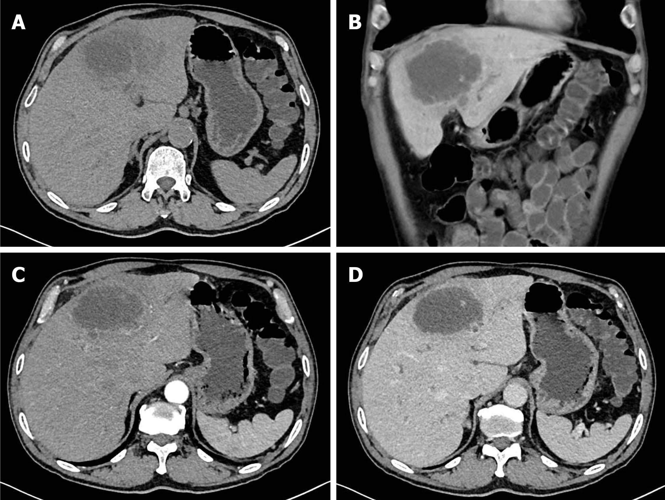
Figure 1 Computed tomography findings.
A and B: Abdominal computed tomography disclosed a low-density mass, 8 cm in diameter, with central necrosis in the left lobe of the liver; C: Enhanced abdominal computed tomography showed a peripheral irregular rim-enhanced mass in the left lobe of the liver without lymph node enlargement; D: Loss of enhancement of the peripheral irregular rim of the mass in the portal phase.
- Citation: Fu RB, Dong XF. Diagnostic challenges and multidisciplinary management of primary adenosquamous carcinoma of the liver: A case report and review of literature. World J Gastrointest Surg 2026; 18(4): 115487
- URL: https://www.wjgnet.com/1948-9366/full/v18/i4/115487.htm
- DOI: https://dx.doi.org/10.4240/wjgs.v18.i4.115487