
















Copyright: ©Author(s) 2026.
World J Gastrointest Surg. Mar 27, 2026; 18(3): 115216
Published online Mar 27, 2026. doi: 10.4240/wjgs.v18.i3.115216
Published online Mar 27, 2026. doi: 10.4240/wjgs.v18.i3.115216
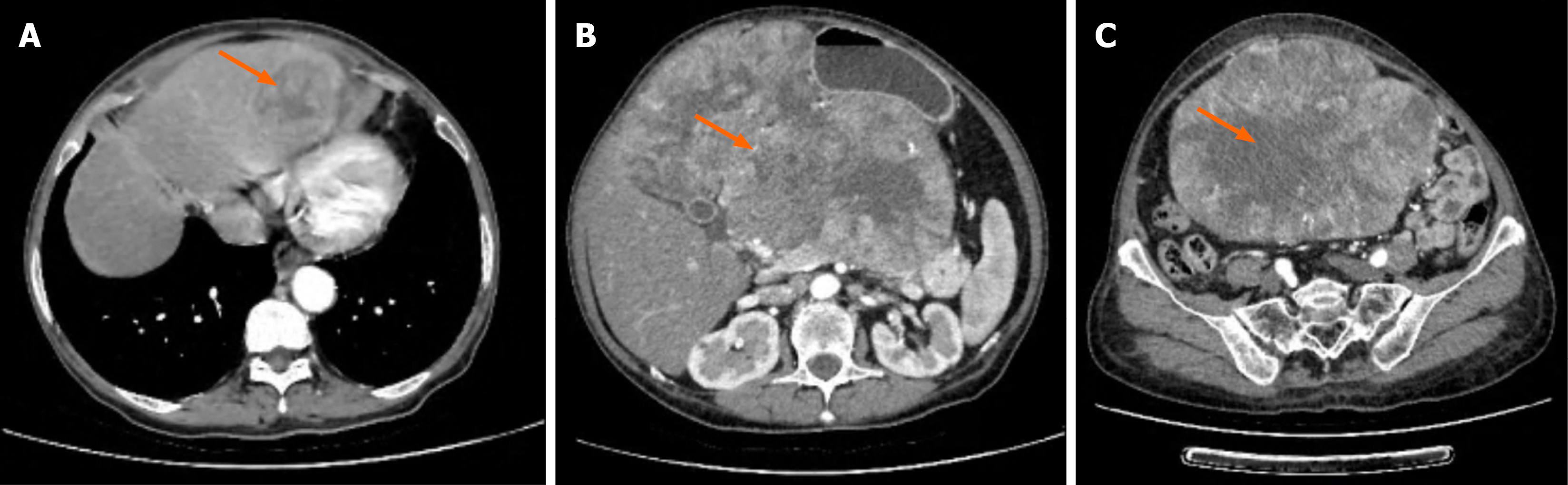
Figure 1 Preoperative contrast-enhanced computed tomography images.
A: Hepatic metastatic lesion (4.4 cm × 3.9 cm); B: Mid-level tumor imaging (orange arrow, 14.1 cm × 18.6 cm × 26.8 cm); C: Tumor extension to the pelvic level. Orange arrow: The tumor mass.
- Citation: Huang K, Wang W, Xu ML, He YS, Zhao JY, Hu XF. Pleomorphic gastric leiomyosarcoma with retroperitoneal extension and hepatic metastasis: A case report. World J Gastrointest Surg 2026; 18(3): 115216
- URL: https://www.wjgnet.com/1948-9366/full/v18/i3/115216.htm
- DOI: https://dx.doi.org/10.4240/wjgs.v18.i3.115216