
















Copyright: ©Author(s) 2026.
World J Gastrointest Surg. Mar 27, 2026; 18(3): 115137
Published online Mar 27, 2026. doi: 10.4240/wjgs.v18.i3.115137
Published online Mar 27, 2026. doi: 10.4240/wjgs.v18.i3.115137
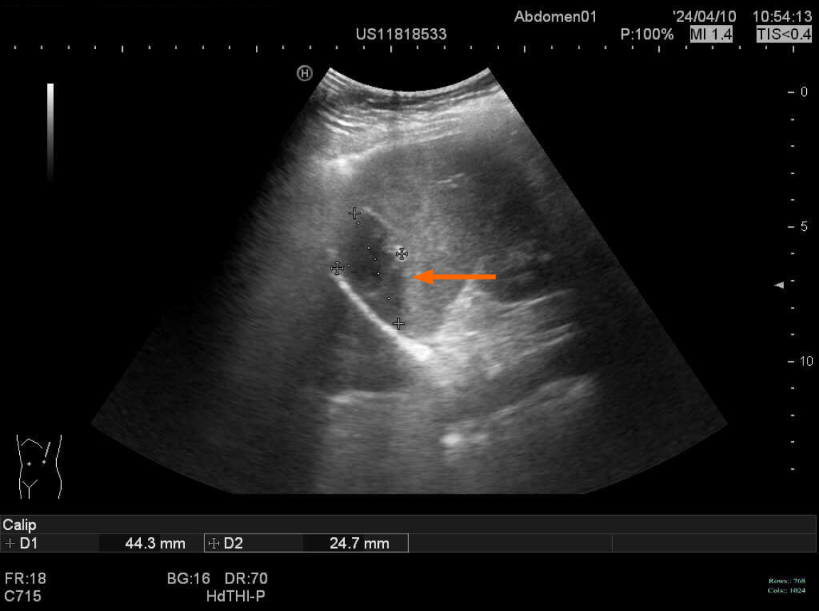
Figure 1 Abdominal ultrasound showing a cystic mass within the spleen.
Orange arrow: A 44 mm × 25 mm hypoechoic area, showing poor internal acoustic transmission and visible by-light bands. No significant blood flow signal detected.
- Citation: Li ZK, Zhang K, Zhang HL, Lv XL, Guo JQ, Tan W. Splenic tuberculous mass following anti-tuberculosis therapy was misdiagnosed as a splenic tumor: A case report. World J Gastrointest Surg 2026; 18(3): 115137
- URL: https://www.wjgnet.com/1948-9366/full/v18/i3/115137.htm
- DOI: https://dx.doi.org/10.4240/wjgs.v18.i3.115137