
















Copyright: ©Author(s) 2026.
World J Gastrointest Surg. Mar 27, 2026; 18(3): 112405
Published online Mar 27, 2026. doi: 10.4240/wjgs.v18.i3.112405
Published online Mar 27, 2026. doi: 10.4240/wjgs.v18.i3.112405
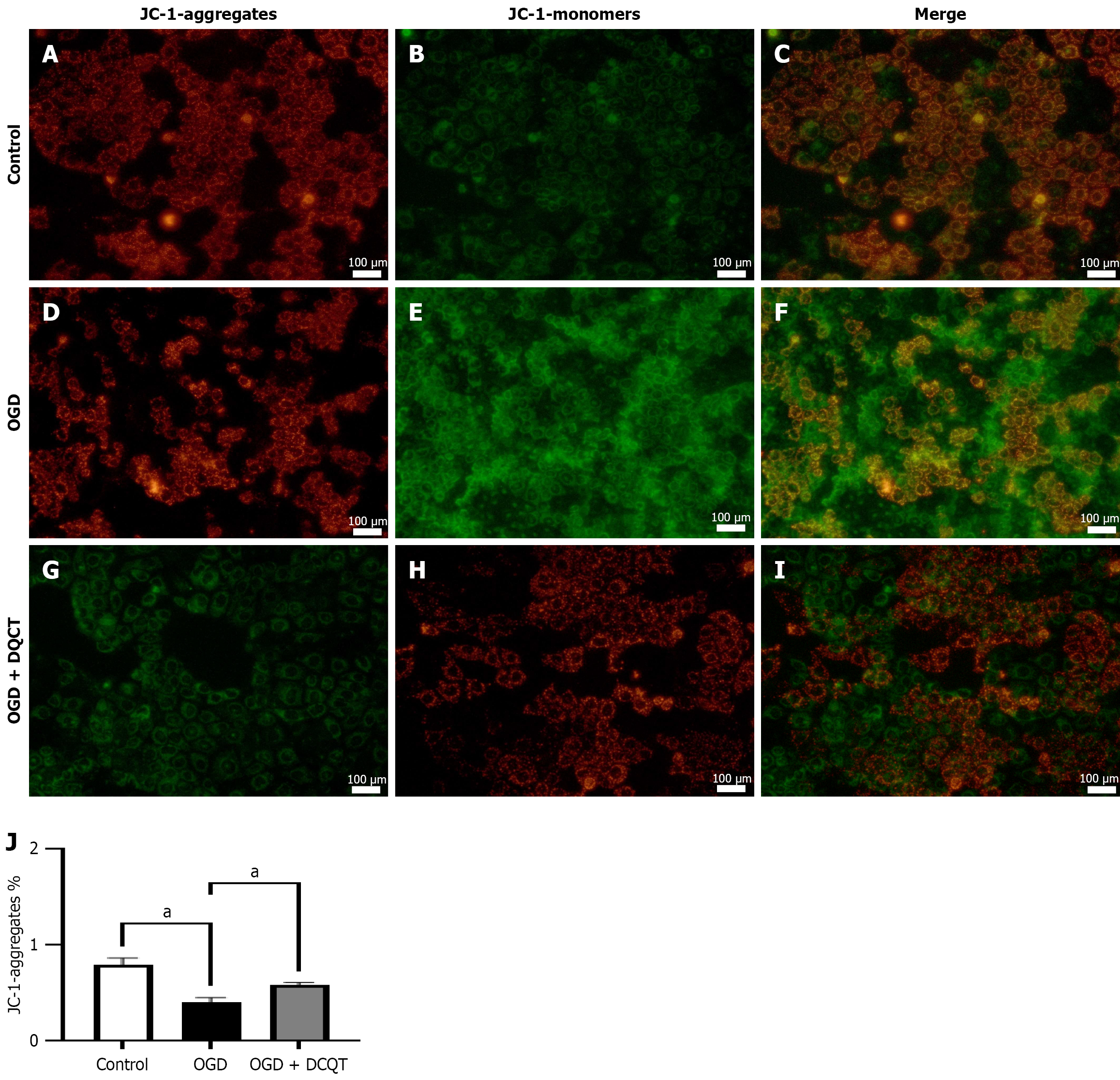
Figure 8 Dachengqitang induces mitochondrial impairment in Caco-2 cells in vitro.
A-C: Mitochondrial membrane potential imaging in Control group after JC-1 staining; D-F: Mitochondrial membrane potential imaging in oxygen-glucose deprivation (OGD) group after JC-1 staining; G-I: Mitochondrial membrane potential imaging in Dachengqitang (DCQT) treatment group after JC-1 staining. Figure showing mitochondrial membrane potential across three groups; J: DCQT protects against OGD-induced mitochondrial membrane potential loss. DCQT treatment significantly reduced JC-1 monomer formation and improved mitochondrial membrane potential compared to OGD treatment alone. aP < 0.05. OGD: Oxygen-glucose deprivation; DCQT: Dachengqitang.
- Citation: Zheng L, Wang J, Xu MQ, Jiang W, Qian L, Ge Y, Feng MK, Zhu YM, Wang MJ, Sun SS, Liu CK, Wang XM, Zhang C. Dachengqitang mitigates endoscopic retrograde cholangiopancreatography-induced pancreatitis. World J Gastrointest Surg 2026; 18(3): 112405
- URL: https://www.wjgnet.com/1948-9366/full/v18/i3/112405.htm
- DOI: https://dx.doi.org/10.4240/wjgs.v18.i3.112405