
















©Author(s) (or their employer(s)) 2026.
World J Gastrointest Surg. Feb 27, 2026; 18(2): 115072
Published online Feb 27, 2026. doi: 10.4240/wjgs.v18.i2.115072
Published online Feb 27, 2026. doi: 10.4240/wjgs.v18.i2.115072
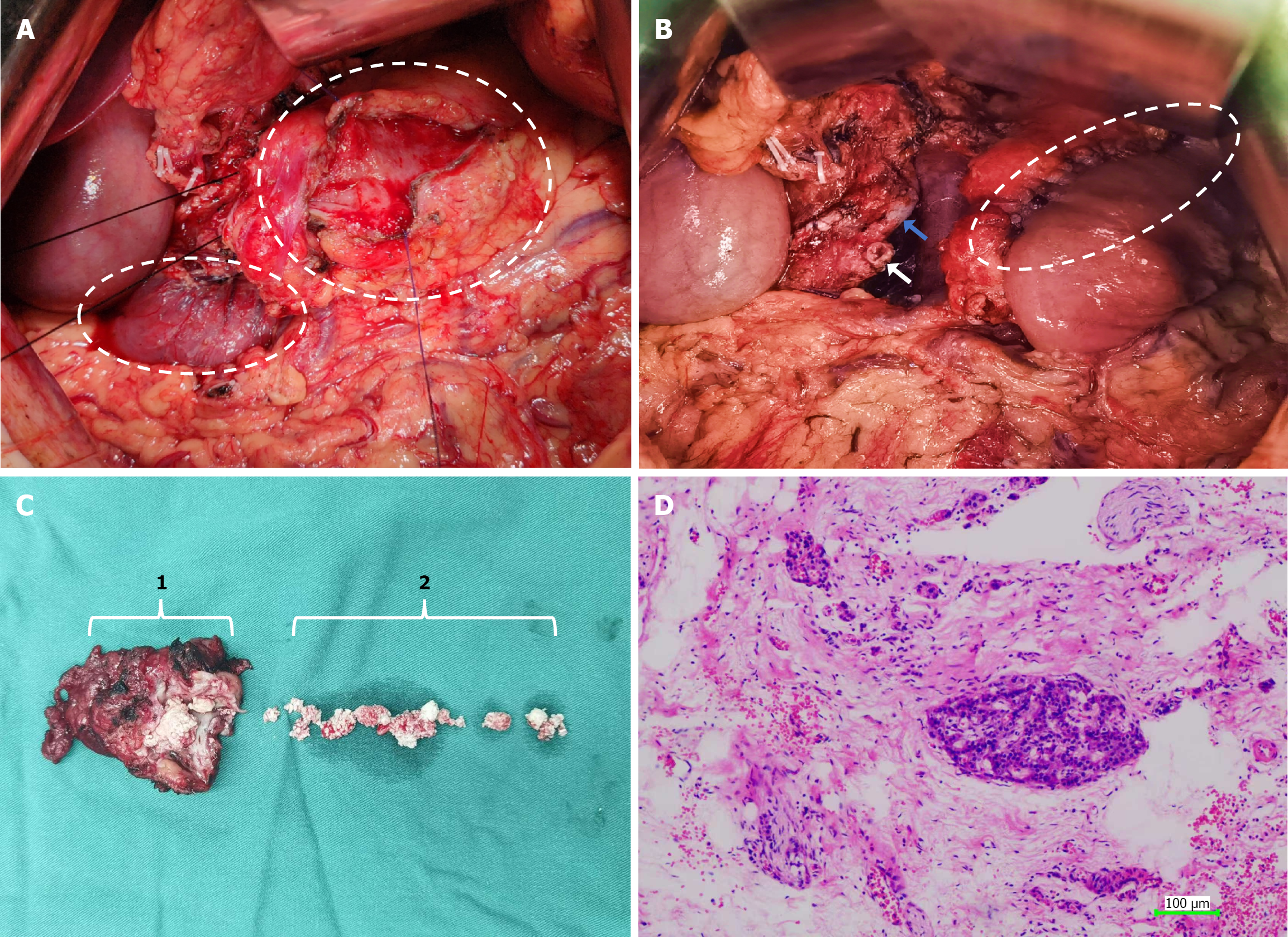
Figure 2 Intraoperative field of view and postoperative specimen.
A: The pancreatic head has been fully resected. As indicated by the large oval, the pancreatic duct in the body and tail has been fully opened longitudinally. The pancreatic parenchyma is atrophic and thin, and the duodenum (indicated by the small oval) is visible below; B: The white oval indicates the pancreatic duct-jejunal anastomosis, with the stump of the main pancreatic duct identifiable on the right side (indicated by the white arrow). The common bile duct remains intact (indicated by the blue arrow) and flows into the duodenum; C: Postoperative specimen. Bracket 1 indicates the intact pancreatic head, and bracket 2 represents the stones removed intraoperatively; D: Pathological hematoxylin and eosin staining of pancreatic head specimen postoperatively.
- Citation: Gou HX, Deng C, Wen Y, Yin ZL, Yang TY, Wang T, Luo H, Cheng L. Pancreatic head resection alongside side-to-side pancreatic duct-jejunostomy for pancreatic stones: A case report and review of literature. World J Gastrointest Surg 2026; 18(2): 115072
- URL: https://www.wjgnet.com/1948-9366/full/v18/i2/115072.htm
- DOI: https://dx.doi.org/10.4240/wjgs.v18.i2.115072