
















©Author(s) (or their employer(s)) 2026.
World J Gastrointest Surg. Feb 27, 2026; 18(2): 115072
Published online Feb 27, 2026. doi: 10.4240/wjgs.v18.i2.115072
Published online Feb 27, 2026. doi: 10.4240/wjgs.v18.i2.115072
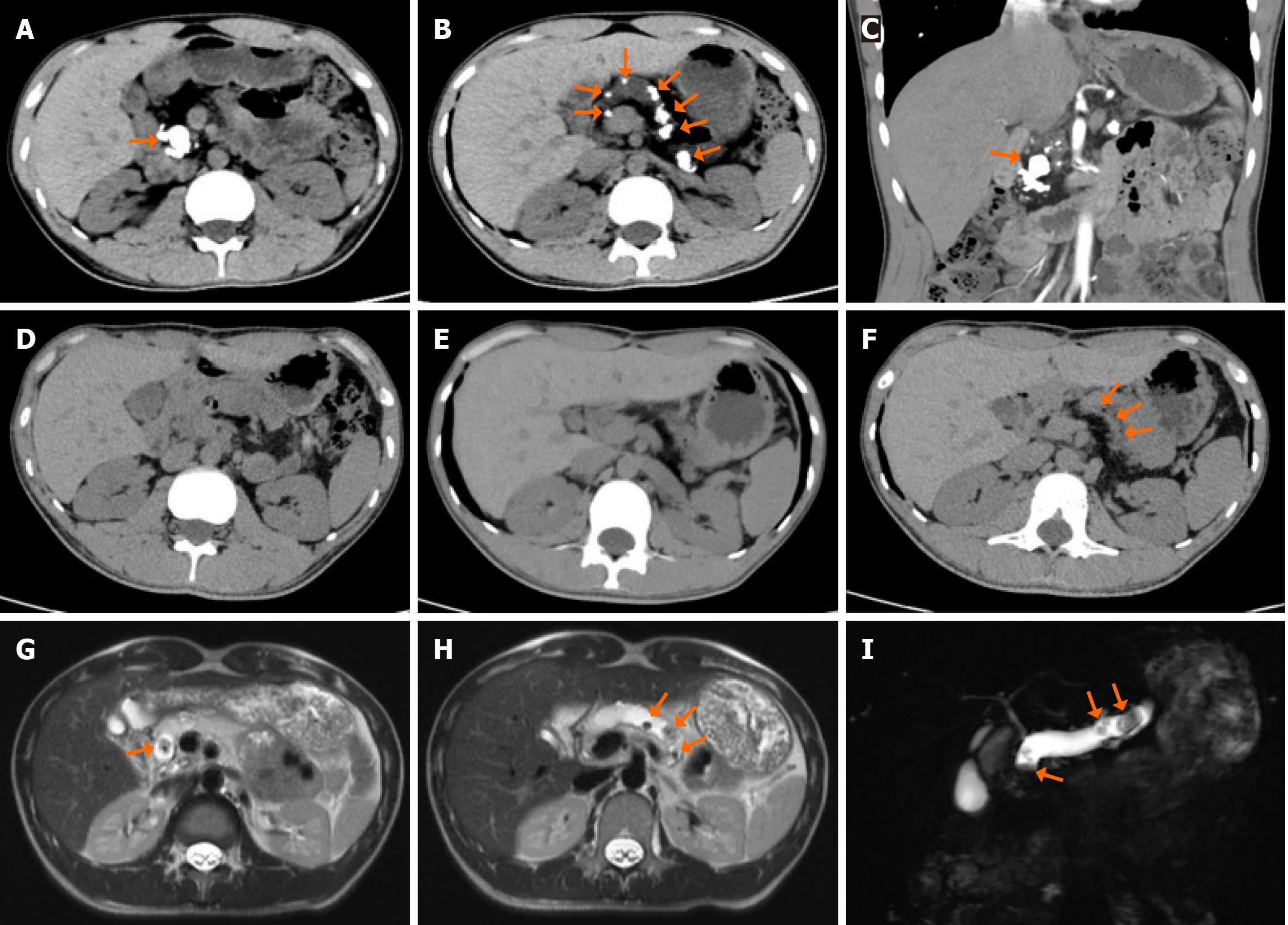
Figure 1 Preoperative and postoperative imaging images.
A: Preoperative plain computed tomography (CT) scan. As indicated by the orange arrows, high-density stones were predominantly located in the pancreatic duct of the pancreatic head; B: Preoperative plain CT scan. As indicated by the orange arrows, multiple high-density stones were scattered in the main pancreatic duct of the pancreatic body and tail; C: Preoperative coronal contrast-enhanced CT scan of the pancreatic head. As indicated by the orange arrows, high-density stones were scattered in the pancreatic duct and pancreatic parenchyma; D: Postoperative plain CT scan of the pancreatic head; E: Postoperative plain CT scan of the pancreatic body and tail; F: Postoperative plain CT scan of the anastomosis. As shown by the orange arrow, no pancreatic duct stones were found at the pancreatic duct-jejunostomy; G: T2-weighted magnetic resonance imaging (MRI) scan of the pancreatic head. As indicated by the orange arrows, a nodular filling defect is observed in the pancreatic duct; H: T2-weighted MRI scan of the pancreatic body. As indicated by the orange arrows, multiple nodular filling defects are observed in the pancreatic duct; I: MRI shows multiple low-intensity signal nodules in the main pancreatic duct.
- Citation: Gou HX, Deng C, Wen Y, Yin ZL, Yang TY, Wang T, Luo H, Cheng L. Pancreatic head resection alongside side-to-side pancreatic duct-jejunostomy for pancreatic stones: A case report and review of literature. World J Gastrointest Surg 2026; 18(2): 115072
- URL: https://www.wjgnet.com/1948-9366/full/v18/i2/115072.htm
- DOI: https://dx.doi.org/10.4240/wjgs.v18.i2.115072