
















©Author(s) (or their employer(s)) 2026.
World J Gastrointest Surg. Feb 27, 2026; 18(2): 114137
Published online Feb 27, 2026. doi: 10.4240/wjgs.v18.i2.114137
Published online Feb 27, 2026. doi: 10.4240/wjgs.v18.i2.114137
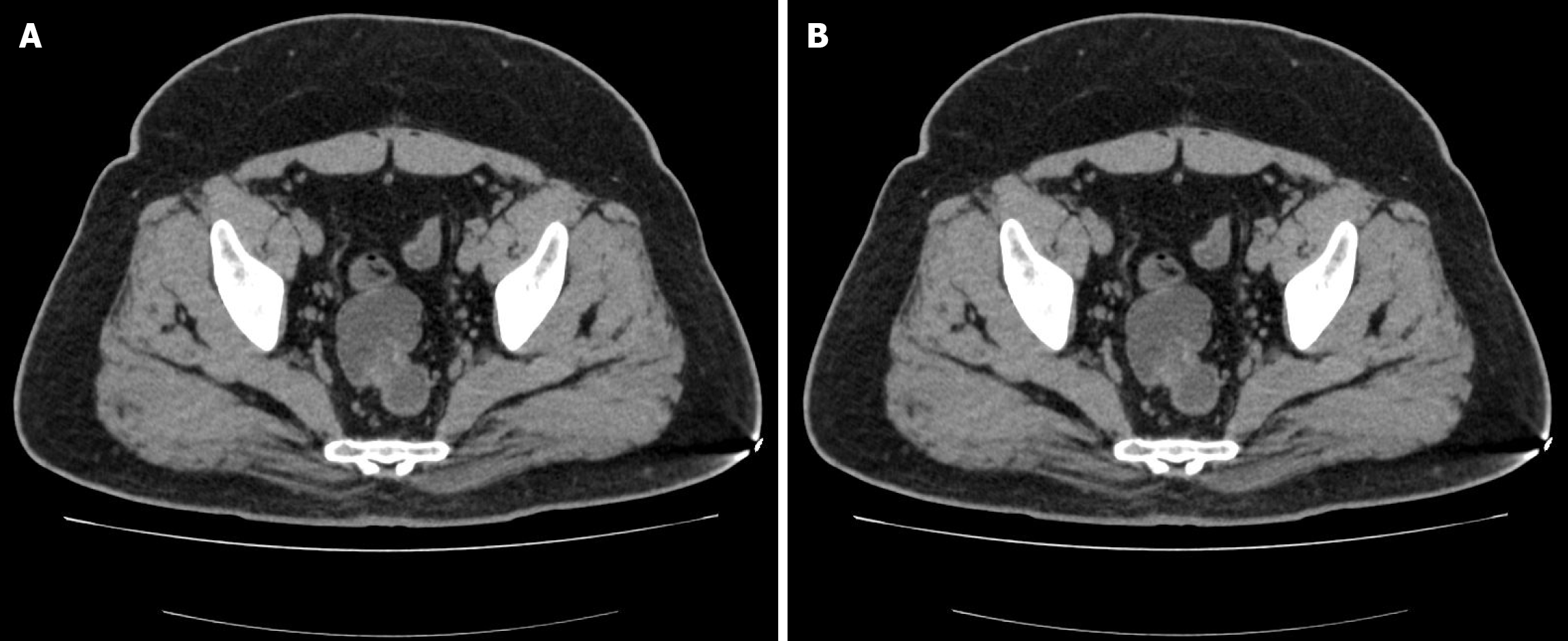
Figure 3 Contrast-enhanced computed tomography.
A: Uneven-density foci on the plain scan, with smooth rectal mucosa and intact intestinal wall; B: Inhomogeneous enhancement is seen in the enhancement phases of the lesion.
- Citation: Zhao T, Jia XR, Li KJ, Zheng WF, Liu XJ. Colitis cystica profunda diagnosed by laparoscopic excision: A case report and review of literature. World J Gastrointest Surg 2026; 18(2): 114137
- URL: https://www.wjgnet.com/1948-9366/full/v18/i2/114137.htm
- DOI: https://dx.doi.org/10.4240/wjgs.v18.i2.114137