
















©Author(s) (or their employer(s)) 2026.
World J Gastrointest Surg. Feb 27, 2026; 18(2): 113698
Published online Feb 27, 2026. doi: 10.4240/wjgs.v18.i2.113698
Published online Feb 27, 2026. doi: 10.4240/wjgs.v18.i2.113698
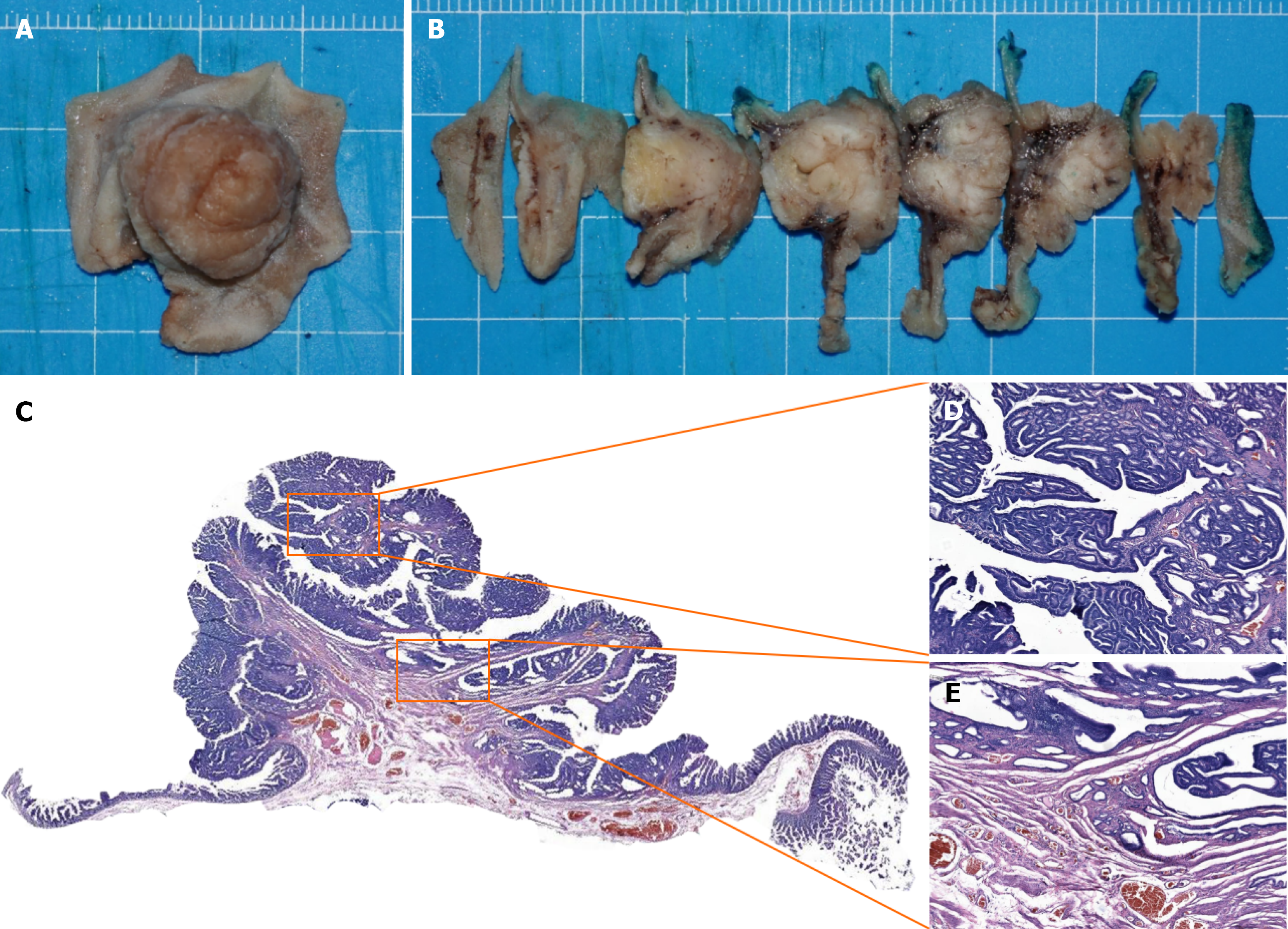
Figure 3 Histopathological evaluation of resected duodenal papillary adenoma.
A: Macroscopic appearance of the formalin-fixed duodenal papillary specimen; B: Representative tissue sections prepared for histopathological examination; C–E: Hematoxylin and eosin-stained sections demonstrating the microscopic architecture of the neoplasms.
- Citation: Xu L, Chen SJ, Wang P, Mao L, Zou XP, Wang L, Wang Y. Safety and efficacy of endoscopic papillectomy for duodenal papillary adenomas: A single-center retrospective study. World J Gastrointest Surg 2026; 18(2): 113698
- URL: https://www.wjgnet.com/1948-9366/full/v18/i2/113698.htm
- DOI: https://dx.doi.org/10.4240/wjgs.v18.i2.113698