
















©Author(s) (or their employer(s)) 2026.
World J Gastrointest Surg. Feb 27, 2026; 18(2): 113698
Published online Feb 27, 2026. doi: 10.4240/wjgs.v18.i2.113698
Published online Feb 27, 2026. doi: 10.4240/wjgs.v18.i2.113698
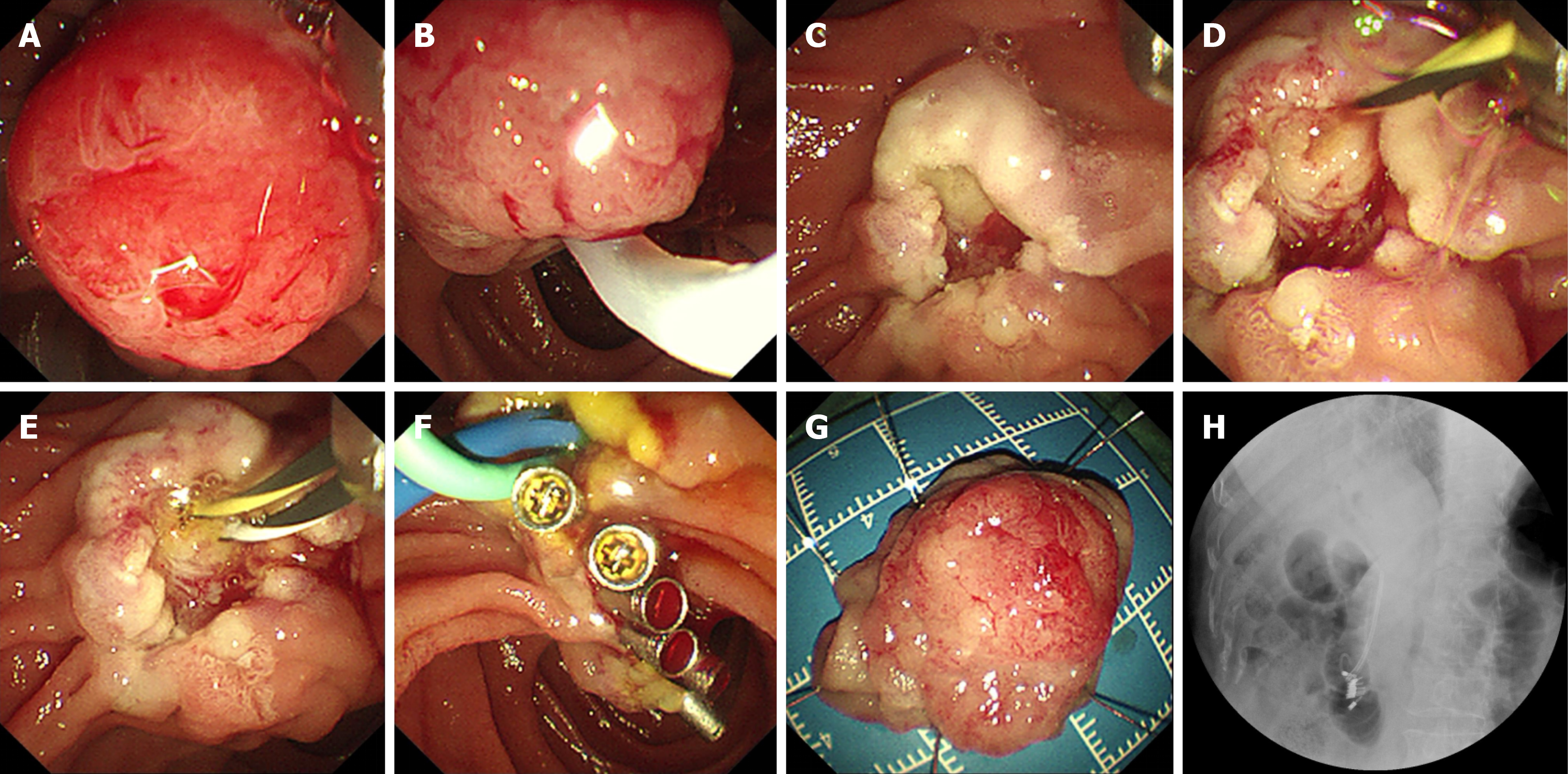
Figure 2 Endoscopic papillectomy for resection of duodenal papillary adenoma.
A: Endoscopic visualization of duodenal papillary adenoma; B: Submucosal injection (e.g., saline with epinephrine) around the lesion to facilitate resection; C and D: En bloc snare excision of the lesion; E and F: Deployment of a 5-Fr, 3-cm pancreatic duct stent to prevent post-procedural pancreatitis, followed by closure of the mucosal defect using hemostatic clips; G: Gross view of the resected specimen; H: X-ray fluoroscopy demonstrating the pancreatic duct stent and titanium clips.
- Citation: Xu L, Chen SJ, Wang P, Mao L, Zou XP, Wang L, Wang Y. Safety and efficacy of endoscopic papillectomy for duodenal papillary adenomas: A single-center retrospective study. World J Gastrointest Surg 2026; 18(2): 113698
- URL: https://www.wjgnet.com/1948-9366/full/v18/i2/113698.htm
- DOI: https://dx.doi.org/10.4240/wjgs.v18.i2.113698