
















Copyright: ©Author(s) 2026.
World J Gastrointest Surg. Nov 27, 2026; 18(11): 125182
Published online Nov 27, 2026. doi: 10.4240/wjgs.125182
Published online Nov 27, 2026. doi: 10.4240/wjgs.125182
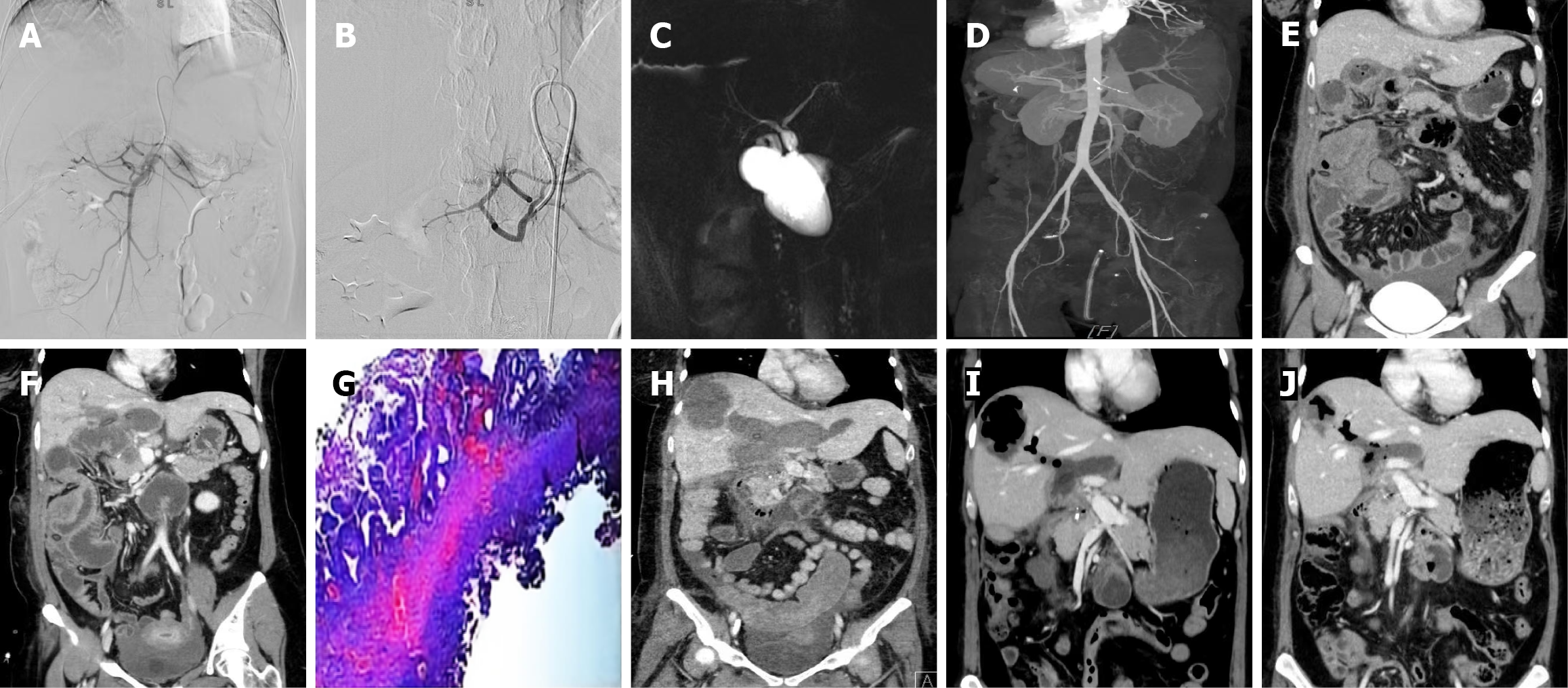
Figure 1 Perioperative imaging examinations.
A and B: Digital subtraction angiography revealing suspected extravasation of the contrast agent at the transitional area of the first branch of the superior mesenteric artery, gastric right artery, and gastric duodenal artery, and preventive embolization was performed; C: Nuclear magnetic resonance imaging revealing cystic dilation of the extrahepatic bile duct; D and E: Computed tomography (CT) angiography and coronal views highlighting ascites and hematoma in the abdominal cavity; F: Coronal views of CT revealing increased ascites and hematoma in the abdominal cavity; G: Postoperative pathology of the common bile duct cyst suggests a benign epithelial cyst of the bile duct alongside microtissue hyperplasia of the cyst wall and scattered chronic inflammatory cell infiltration, consistent with the diagnosis of bile duct cyst; H: Coronary views of CT indicating the formation of liver abscess and pelvic effusion; I: Coronary views of CT indicating a reduction in the abscess cavity and a decrease in the pus volume of the liver abscess; J: Coronary views of CT indicating that the abscess cavity of the liver abscess has basically healed.
- Citation: Yuan SR, Cao L, Li XJ, Cao Y. Acquired hemophilia A following choledochal cyst excision - highlighting postoperative bile leak and infection as potential triggers: A case report. World J Gastrointest Surg 2026; 18(11): 125182
- URL: https://www.wjgnet.com/1948-9366/full/v18/i11/125182.htm
- DOI: https://dx.doi.org/10.4240/wjgs.125182