
















Copyright: ©Author(s) 2026.
World J Gastrointest Surg. Nov 27, 2026; 18(11): 124759
Published online Nov 27, 2026. doi: 10.4240/wjgs.124759
Published online Nov 27, 2026. doi: 10.4240/wjgs.124759
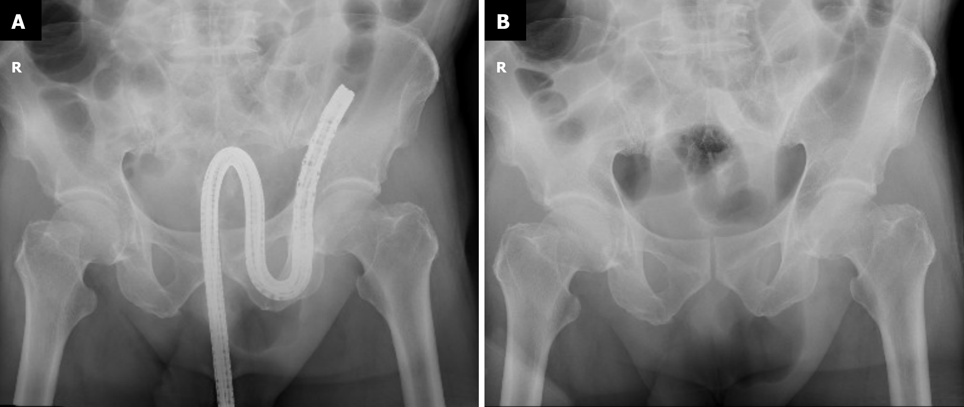
Figure 1 Abdominal X-ray.
A: Pelvic X-ray showing the colonoscope stuck in a reverse “S” configuration in the left pelvic cavity; B: Successful withdrawal of the colonoscope following manual reduction of the inguinal hernia.
- Citation: Yao WX, Li YH, Deng B, Wu LG, Du L. Colonoscope incarceration in an inguinal hernia complicated by ischemic bowel disease: A case report. World J Gastrointest Surg 2026; 18(11): 124759
- URL: https://www.wjgnet.com/1948-9366/full/v18/i11/124759.htm
- DOI: https://dx.doi.org/10.4240/wjgs.124759