
















Copyright: ©Author(s) 2026.
World J Gastrointest Surg. Oct 27, 2026; 18(10): 123763
Published online Oct 27, 2026. doi: 10.4240/wjgs.123763
Published online Oct 27, 2026. doi: 10.4240/wjgs.123763
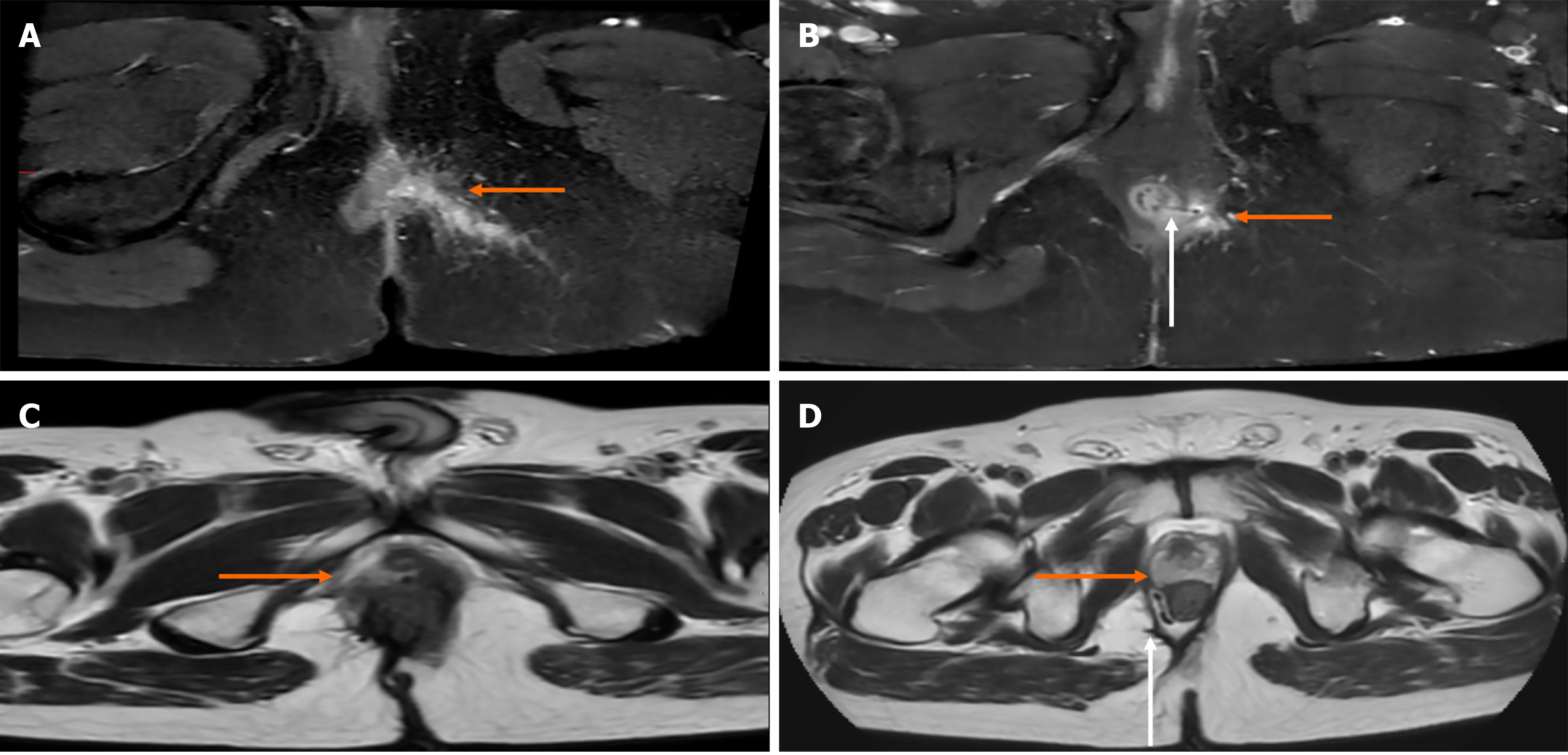
Figure 4 Pre- and post-seton pelvic magnetic resonance imaging illustrating abscess cavity evolution in complex anal fistulas.
A: Preoperative pelvic magnetic resonance imaging (MRI) showing an abscess cavity (orange arrow); B: Follow-up MRI showing resolution of the abscess cavity (orange arrow) after seton placement, and that the loose seton remained visible (white arrow); C: Preoperative pelvic MRI in a different patient showing an abscess cavity (orange arrow); D: Same MRI as in (C) showing a persistent abscess and sinus tract (orange arrow) despite ongoing drainage through a loose seton (white arrow).
- Citation: Edizsoy A, Tahirli N, Tanyeri A, Tunçyürek P. Timing of ligation of the intersphincteric fistula tract after loose seton drainage in complex anal fistulas. World J Gastrointest Surg 2026; 18(10): 123763
- URL: https://www.wjgnet.com/1948-9366/full/v18/i10/123763.htm
- DOI: https://dx.doi.org/10.4240/wjgs.123763