
















Copyright: ©Author(s) 2026.
World J Gastrointest Surg. Oct 27, 2026; 18(10): 123695
Published online Oct 27, 2026. doi: 10.4240/wjgs.123695
Published online Oct 27, 2026. doi: 10.4240/wjgs.123695
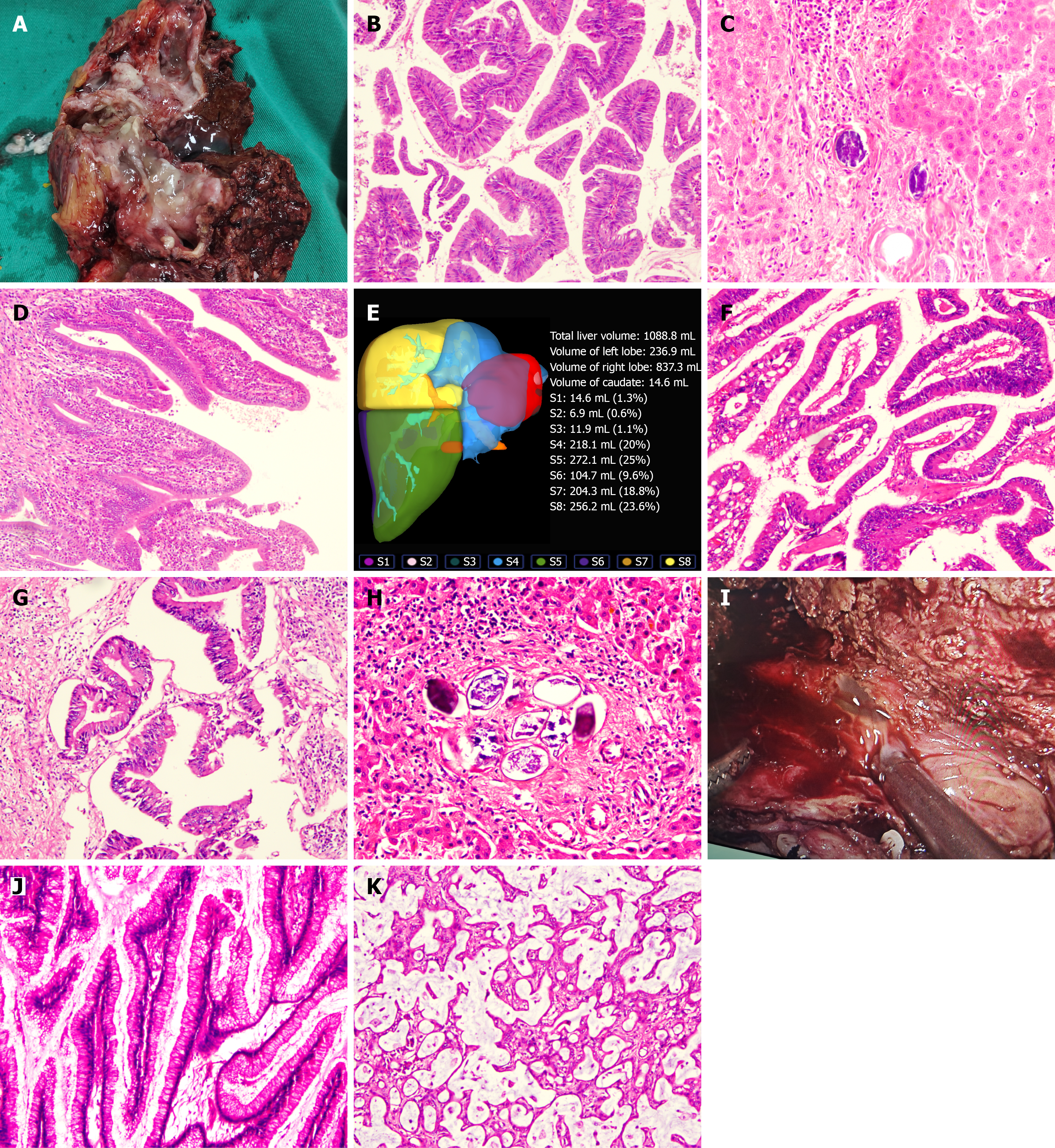
Figure 2 Gross specimen, pathology of patients.
A: Gross specimen of patient 1 showing markedly dilated bile ducts filled with mucin and a papillary mass; pathology of patient 1 (B-D); B: Papillary architecture with fibrovascular cores on histopathological examination [hematoxylin and eosin (HE) × 100]; C: Schi
- Citation: Yang YL, Zhang TQ, Le WJ, Zhou L, Cheng H, Li YJ, Zhang KM, Yang ZC, Peng T, Lu HF. Debulking surgery for non-radically resectable intraductal papillary neoplasm of the bile duct: Three case reports and literature review. World J Gastrointest Surg 2026; 18(10): 123695
- URL: https://www.wjgnet.com/1948-9366/full/v18/i10/123695.htm
- DOI: https://dx.doi.org/10.4240/wjgs.123695