
















Copyright: ©Author(s) 2026.
World J Gastrointest Surg. Oct 27, 2026; 18(10): 123208
Published online Oct 27, 2026. doi: 10.4240/wjgs.123208
Published online Oct 27, 2026. doi: 10.4240/wjgs.123208
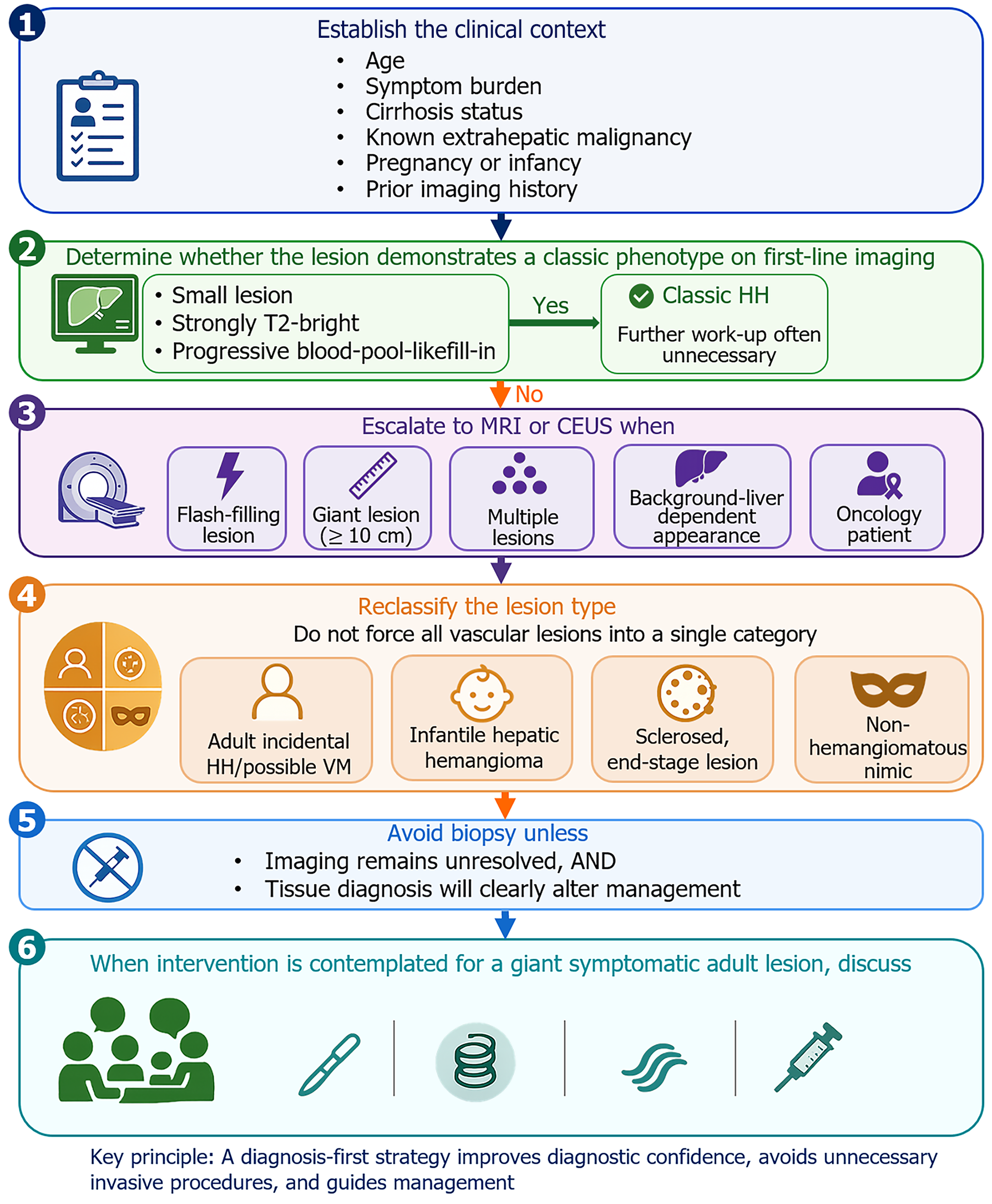
Figure 2 A practical diagnosis-first framework for hepatic hemangioma.
The flowchart illustrates a six-step multidisciplinary diagnostic and management strategy. The framework highlights the importance of clinical context (Step 1), specific indications for escalation to advanced multimodality imaging (Steps 2-3), the necessity of reclassifying lesion types (Step 4), stringent criteria for biopsy (Step 5), and customized interventional or surgical management based on lesion biology (Step 6). HH: Hepatic hemangioma; MRI: Magnetic resonance imaging; CEUS: Contrast-enhanced ultrasound; VM: Venous malformation.
- Citation: Ma YX, Zhou CM, Yilihaer Y, Jiang TM, Ruze R, Shao YM, Wen H, Aji T. Diagnosis and surgical decision-making in hepatic hemangioma: From venous malformation overlap to artificial intelligence-assisted multimodality imaging. World J Gastrointest Surg 2026; 18(10): 123208
- URL: https://www.wjgnet.com/1948-9366/full/v18/i10/123208.htm
- DOI: https://dx.doi.org/10.4240/wjgs.123208