
















Copyright: ©Author(s) 2026.
World J Gastrointest Surg. Oct 27, 2026; 18(10): 123199
Published online Oct 27, 2026. doi: 10.4240/wjgs.123199
Published online Oct 27, 2026. doi: 10.4240/wjgs.123199
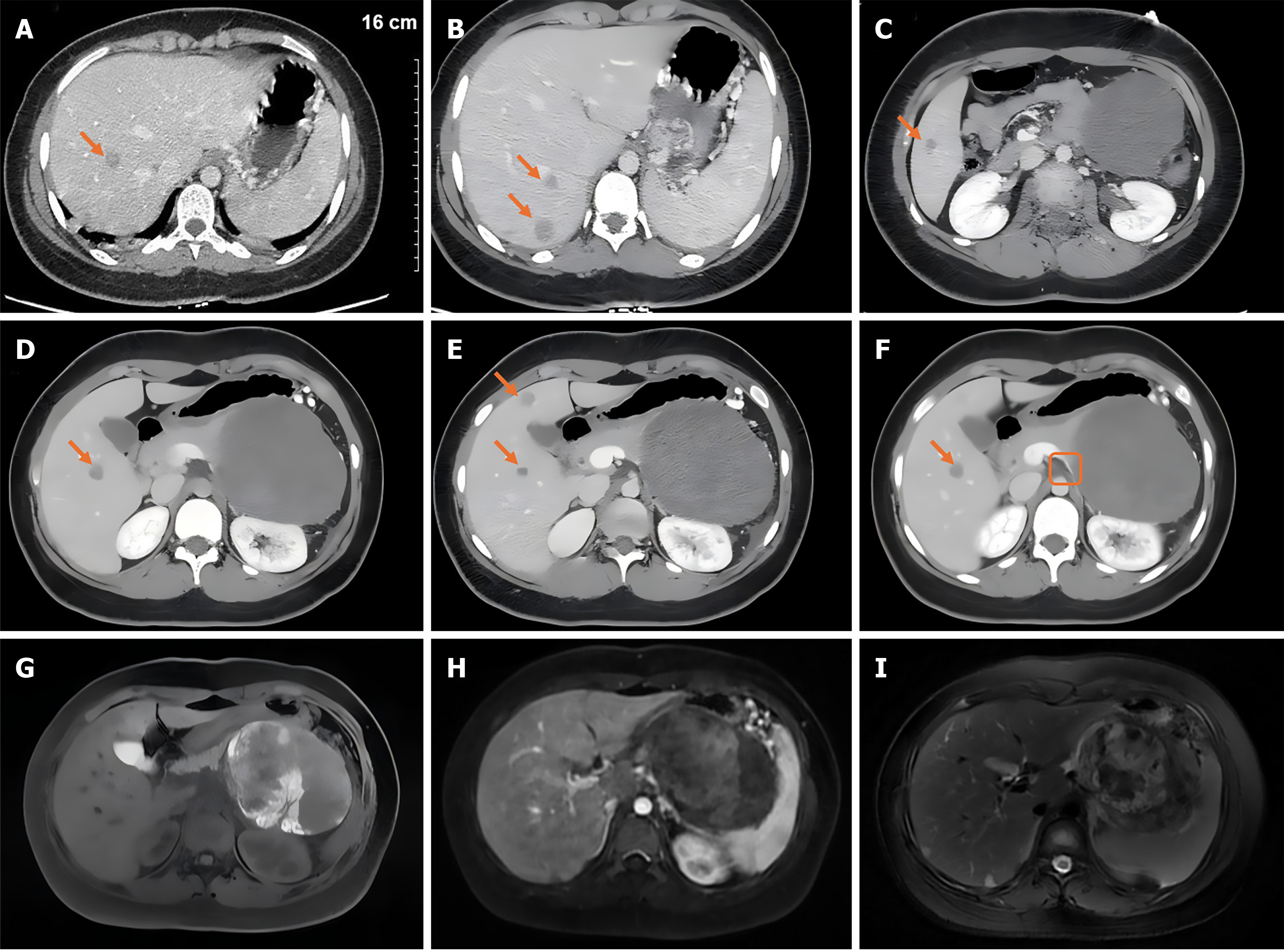
Figure 1 Contrast-enhanced abdominal computed tomography/magnetic resonance imaging.
A-F: Multiple metastatic lesions are demonstrated in hepatic segments VIII, VII, VI, V, IV (arrows); F: The portal venous phase of contrast-enhanced computed tomography shows compression and narrowing of the splenic vein; G: Fat-suppressed magnetic resonance imaging (MRI) images reveal intratumoral hemorrhage; H: T1-weighted contrast-enhanced MRI demonstrates heterogeneous enhancement within the tumor; I: T2-weighted MRI shows mixed cystic-solid signals. All images are displayed at an identical scale (scale bar in A: 1 cm per division, total 16 cm).
- Citation: Zhou XY, Chen T, Liang J, Yue JQ, Wu LL, Li HM, Liu ZH, Peng CJ, Liu GW. Aggressive pediatric solid pseudopapillary neoplasms of the pancreas presented as acute abdomen: A case report and review of literature. World J Gastrointest Surg 2026; 18(10): 123199
- URL: https://www.wjgnet.com/1948-9366/full/v18/i10/123199.htm
- DOI: https://dx.doi.org/10.4240/wjgs.123199