
















Copyright: ©Author(s) 2026.
World J Gastrointest Surg. Oct 27, 2026; 18(10): 123093
Published online Oct 27, 2026. doi: 10.4240/wjgs.123093
Published online Oct 27, 2026. doi: 10.4240/wjgs.123093
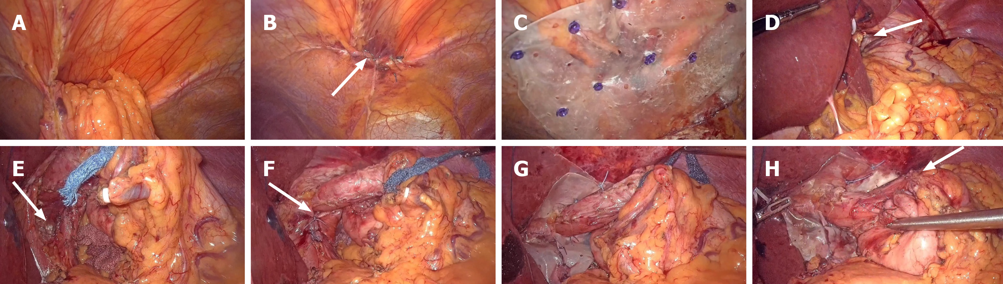
Figure 2 Intraoperative laparoscopic findings and key operative steps.
A: Laparoscopic exploration revealing a retrosternal right-sided diaphragmatic defect with the greater omentum as the hernia content; B: Primary suture closure of the Morgagni hernia defect; C: Fixation of the biological mesh for defect augmentation; D: Laparoscopic visualization of the sliding hiatus hernia; E: Complete reduction of the herniated gastric fundus into the abdominal cavity with full mobilization of the gastric fundus and distal esophagus; F: Primary suture reconstruction of the bilateral diaphragmatic crura; G: Augmentation repair of the esophageal hiatus with biological mesh; H: 270-degree “U” shaped Toupet fundoplication around the gastroesophageal junction.
- Citation: Xia ZL, Luo KC, Tang J, Sun L. Laparoscopic one-stage repair of concurrent Morgagni hernia and hiatus hernia: A case report and review of literature. World J Gastrointest Surg 2026; 18(10): 123093
- URL: https://www.wjgnet.com/1948-9366/full/v18/i10/123093.htm
- DOI: https://dx.doi.org/10.4240/wjgs.123093