
















Copyright: ©Author(s) 2026.
World J Gastrointest Surg. Oct 27, 2026; 18(10): 123036
Published online Oct 27, 2026. doi: 10.4240/wjgs.123036
Published online Oct 27, 2026. doi: 10.4240/wjgs.123036
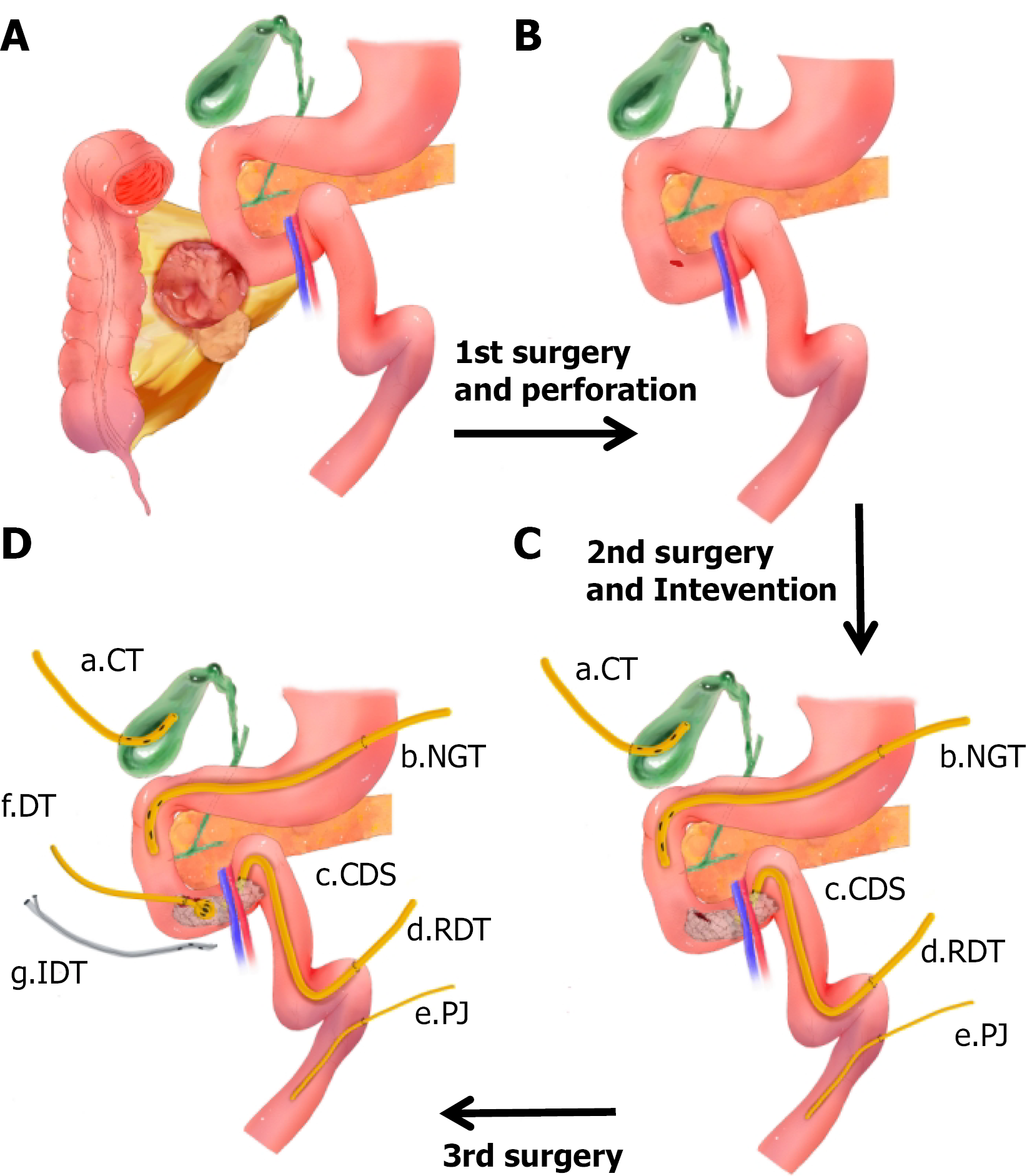
Figure 2 Surgical illustrations.
A and B: First operation: Multiple soft tissue tumors of the colonic mesentery and the right hemicolon were resected. The larger tumor was tightly adherent to the lateral wall of the duodenum; B and C: Second operation and intervention: Intraoperative exploration revealed a perforation on the anterior wall of the horizontal portion of the duodenum. The perforation was repaired with sutures and reinforced using a ligamentum teres hepatis patch. Gastroenteric and biliary diversions were created, and a feeding jejunostomy tube was placed. Postoperative examination indicated persistent unhealed duodenal perforation, and a covered duodenal stent was placed over the perforation site; C and D: Third operation: A duodenostomy tube was directly placed at the perforation site and secured with a wrap of the ligamentum teres hepatis. Additionally, a paraduodenal drain with irrigation and suction capabilities was placed. CT: Cholecystostomy tube; NT: Nasogastric tube; CDS: Ccovered duodenal stent; RDT: Retrograde duodenal decompression tube; PJ: Percutaneous jejunostomy tube; DT: Duodenostomy tube; IDT: Irrigable drainage tube.
- Citation: Dong LB, Wu MW, Han JS, Lin C, Wang WB. Multidisciplinary management of postoperative duodenal perforation using multiple drainage and stenting: A case report and review of literature. World J Gastrointest Surg 2026; 18(10): 123036
- URL: https://www.wjgnet.com/1948-9366/full/v18/i10/123036.htm
- DOI: https://dx.doi.org/10.4240/wjgs.123036