
















Copyright: ©Author(s) 2026.
World J Gastrointest Surg. Oct 27, 2026; 18(10): 122889
Published online Oct 27, 2026. doi: 10.4240/wjgs.122889
Published online Oct 27, 2026. doi: 10.4240/wjgs.122889
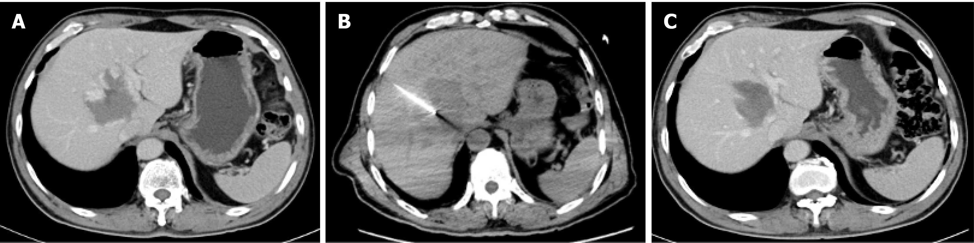
Figure 3 Percutaneous computed tomography-guided radiofrequency ablation of a 6.
5 cm hepatic paracaval portion hemangioma. A: Preoperative computed tomography (CT) scan demonstrated a hepatic caudate lobe hemangioma of 6.5 cm in paracaval portion of the caudate lobe; B: CT image showing radiofrequency needle within the lesion, lesion approached through the right intercostal approach; C: Follow-up CT at 1 month showed that the hemangioma was significantly smaller than before and had no enhancement.
- Citation: Xu L, Gao RZ, Dong SY, Kong J, Yao CY, Wu SL, Ke S, Zhou ZT, Gao J, Wang SH. Efficacy and safety of thermal ablation for large hepatic caudate lobe hemangioma. World J Gastrointest Surg 2026; 18(10): 122889
- URL: https://www.wjgnet.com/1948-9366/full/v18/i10/122889.htm
- DOI: https://dx.doi.org/10.4240/wjgs.122889