
















Copyright: ©Author(s) 2026.
World J Gastrointest Surg. Oct 27, 2026; 18(10): 122889
Published online Oct 27, 2026. doi: 10.4240/wjgs.122889
Published online Oct 27, 2026. doi: 10.4240/wjgs.122889
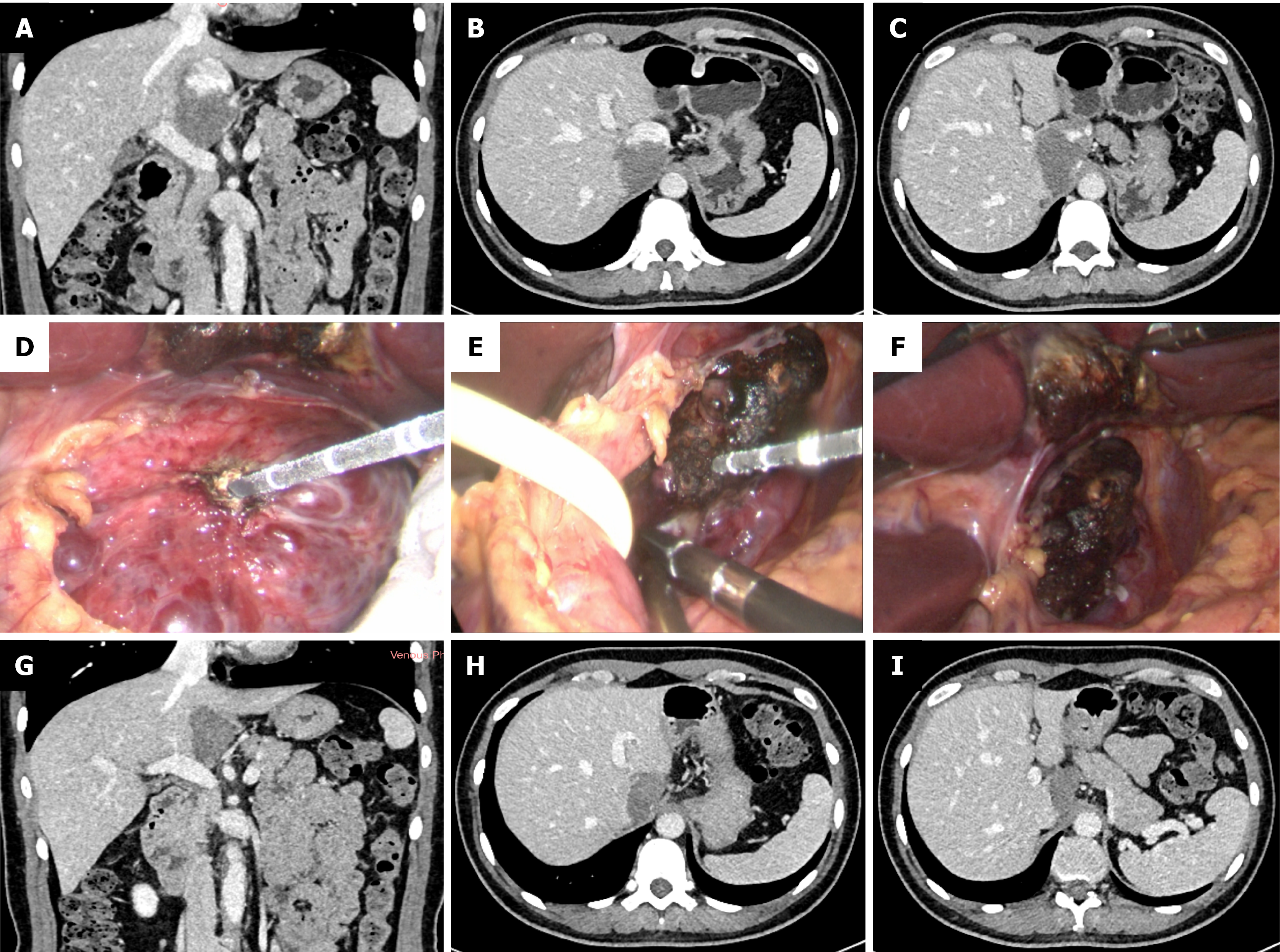
Figure 1 Microwave ablation of a 6.
4 cm hepatic Spiegel lobe hemangioma. A-C: Preoperative computed tomography (CT) scan demonstrated a 6.4 cm hepatic caudate lobe hemangioma (Spiegel lobe); D-F: Expose the hepatic hemangioma and perform microwave ablation; G-I: Follow-up abdominal contrast-enhanced CT at 3 months postoperatively.
- Citation: Xu L, Gao RZ, Dong SY, Kong J, Yao CY, Wu SL, Ke S, Zhou ZT, Gao J, Wang SH. Efficacy and safety of thermal ablation for large hepatic caudate lobe hemangioma. World J Gastrointest Surg 2026; 18(10): 122889
- URL: https://www.wjgnet.com/1948-9366/full/v18/i10/122889.htm
- DOI: https://dx.doi.org/10.4240/wjgs.122889