
















Copyright: ©Author(s) 2026.
World J Gastrointest Surg. Oct 27, 2026; 18(10): 122861
Published online Oct 27, 2026. doi: 10.4240/wjgs.122861
Published online Oct 27, 2026. doi: 10.4240/wjgs.122861
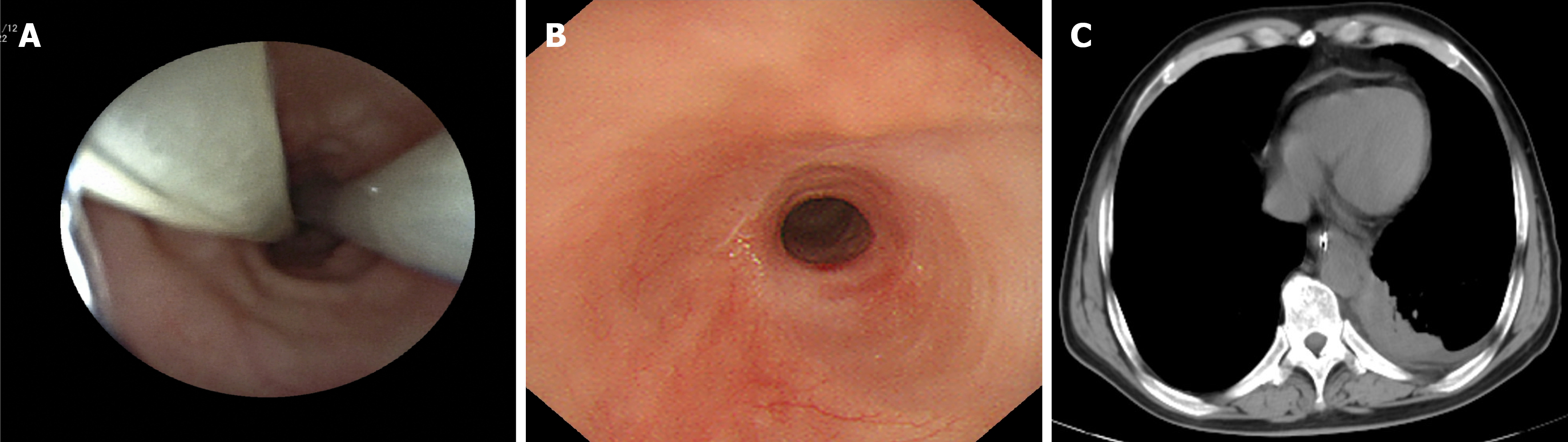
Figure 3 Follow-up examination after one week of drainage and images showing fistula healing.
A: Gastroscopy at the 2-week follow-up showing a well-positioned esophageal drainage tube; B: Gastroscopy on January 25, 2023 showing scar formation at the fistula site, confirming healing; C: Repeat chest computed tomography showing no significant effusion or pneumothorax, with pleural effusion largely absorbed.
- Citation: Ding WJ, Tang HZ, Zhou XR, Liu WH, Xu GR. Conservative management of a delayed esophagomediastinal fistula after endoscopic submucosal dissection: A case report. World J Gastrointest Surg 2026; 18(10): 122861
- URL: https://www.wjgnet.com/1948-9366/full/v18/i10/122861.htm
- DOI: https://dx.doi.org/10.4240/wjgs.122861