
















©The Author(s) 2026.
World J Gastrointest Surg. Jan 27, 2026; 18(1): 114041
Published online Jan 27, 2026. doi: 10.4240/wjgs.v18.i1.114041
Published online Jan 27, 2026. doi: 10.4240/wjgs.v18.i1.114041
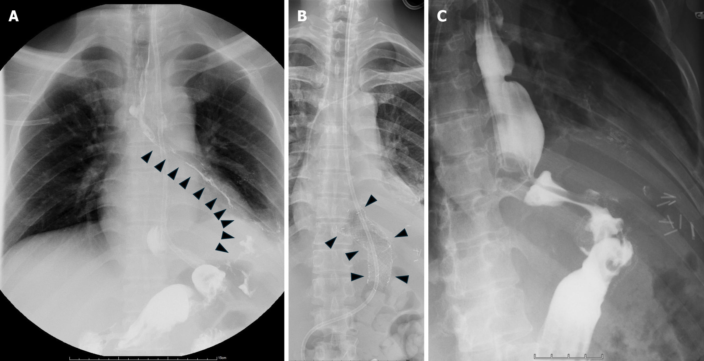
Figure 3 Treatment imaging.
A: Upper gastrointestinal series revealed a left-sided gastrobronchial fistula (arrowhead); B: Chest X-ray demonstrated the proper position of the covered self-expandable metallic stent (arrowhead); C: Upper gastrointestinal series after stent removal showed no contrast leak into the pleural cavity.
- Citation: Fang KH, Chang HM, Wu TH. Successful treatment of gastrobronchial fistula following laparoscopic sleeve gastrectomy: A case report and review of literature. World J Gastrointest Surg 2026; 18(1): 114041
- URL: https://www.wjgnet.com/1948-9366/full/v18/i1/114041.htm
- DOI: https://dx.doi.org/10.4240/wjgs.v18.i1.114041