
















©The Author(s) 2026.
World J Gastrointest Surg. Jan 27, 2026; 18(1): 114022
Published online Jan 27, 2026. doi: 10.4240/wjgs.v18.i1.114022
Published online Jan 27, 2026. doi: 10.4240/wjgs.v18.i1.114022
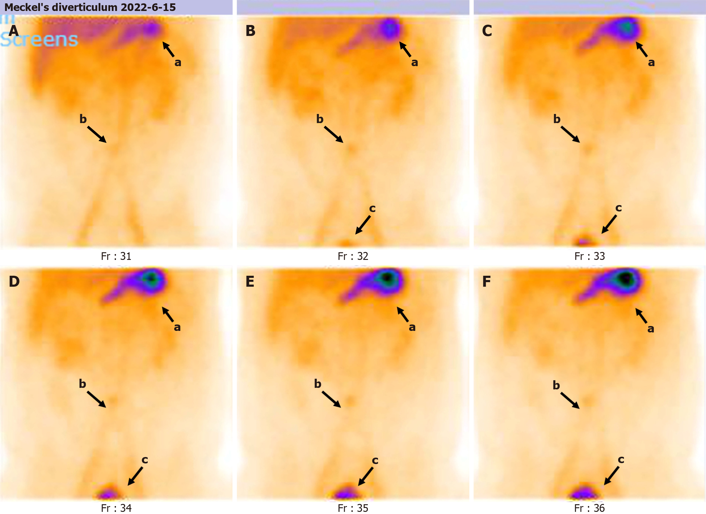
Figure 1 Pre-operative technetium-99m pertechnetate Meckel scan.
After intravenous administration of the radiotracer (99mTcO4-), dynamic abdominal imaging was performed for 30 minutes, with one frame acquired every 5 minutes, resulting in six frames. A: Immediate gastric visualization is observed (position a in the illustration). A circular lesion with mildly increased tracer uptake is detected in the mid-abdomen (position b); B: Approximately 10 minutes post-injection, the bladder is visualized (position c), while positions a and b remain fixed with progressive accumulation of tracer; C-F: Over time, tracer uptake within the lesion is slightly increased, without notable changes in morphology or location.
- Citation: Yu ZH, Ling CR, Yu JY, Zhang QH, Wei SS. Long-term disease-free survival following preoperative diagnosis and laparoscopic radical resection for Meckel’s diverticulum adenocarcinoma: A case report. World J Gastrointest Surg 2026; 18(1): 114022
- URL: https://www.wjgnet.com/1948-9366/full/v18/i1/114022.htm
- DOI: https://dx.doi.org/10.4240/wjgs.v18.i1.114022