
















©The Author(s) 2026.
World J Gastrointest Surg. Jan 27, 2026; 18(1): 113894
Published online Jan 27, 2026. doi: 10.4240/wjgs.v18.i1.113894
Published online Jan 27, 2026. doi: 10.4240/wjgs.v18.i1.113894
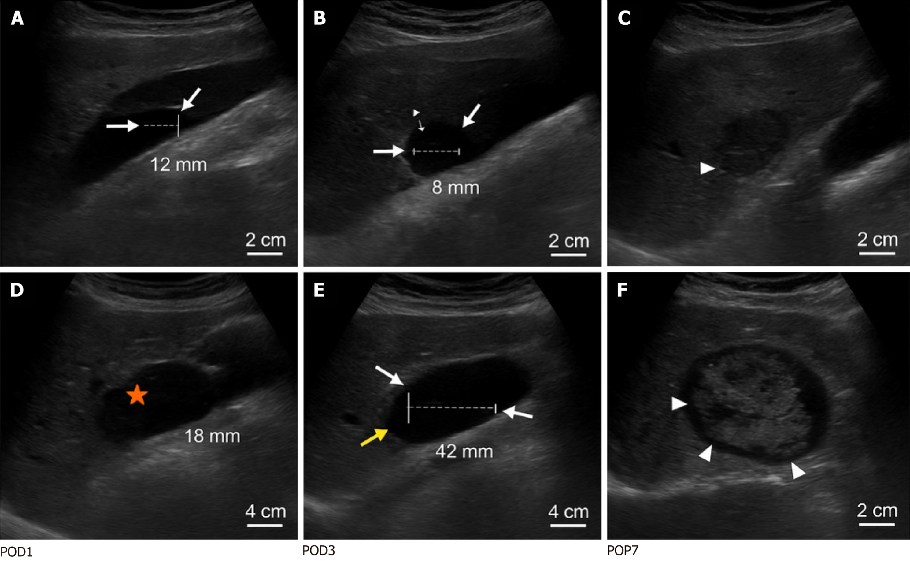
Figure 1 Imaging examination of typical case.
A-C: Detection of a 58-year-old man after distal gastrectomy. Postoperative day (POD1): Small anechoic collection (arrow) in the hepatic and renal recess (Morison’s capsule), depth = 12 mm, volume approximately 15 mL, C-reactive protein (CRP) = 28 mg/L (A). POD3: Reduction in collection (depth = 8 mm, arrow) with silencing mode maintained. CRP = 32 mg/L (B). POD7: Complete resolution of the previously observed fluid collection. The arrow indicates the absence of any residual anechoic area, consistent with full radiological resolution complete resolution CRP = 12 mg/L (C); D-F: A 63-year-old man with mixed collections of infection and progression to an abscess after total gastrectomy. POD1: Heterogeneous collection of subtle internal echoes (stars) in the splenic bed, depth = 18 mm, volume approximately 35 mL, CRP = 46 mg/L, orange star is internal echo/fragment (D). POD3: Dilated (depth = 42 mm) with a septum (arrow) and echo enhancement. CRP = 89 mg/L (above the diagnostic threshold), yellow arrow is partition/wall thickening (E). POD7: Complex mass with wall thickening (arrow) and internal debris, confirmed as an Escherichia coli abscess by computed tomography-guided drainage (draining 210 mL of pus). CRP = 112 mg/L (F). POD: Postoperative day.
- Citation: Gao XX, Su SH, Huang DD. Dynamic ultrasound monitoring of intraperitoneal effusion for predicting complications after laparoscopic gastrectomy. World J Gastrointest Surg 2026; 18(1): 113894
- URL: https://www.wjgnet.com/1948-9366/full/v18/i1/113894.htm
- DOI: https://dx.doi.org/10.4240/wjgs.v18.i1.113894