
















©The Author(s) 2025.
World J Gastrointest Surg. Sep 27, 2025; 17(9): 110796
Published online Sep 27, 2025. doi: 10.4240/wjgs.v17.i9.110796
Published online Sep 27, 2025. doi: 10.4240/wjgs.v17.i9.110796
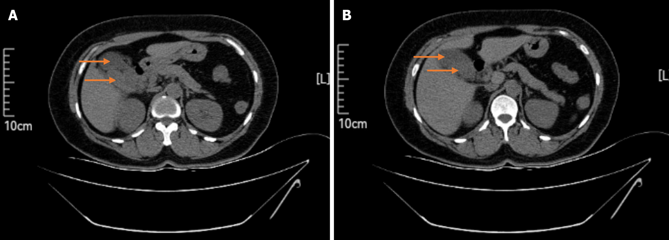
Figure 2 Computed tomography scan showing a duplicated gallbladder.
No gallstones are discernable on the tomographic images. A and B: The different layers of the computed tomography scan. The orange arrows indicate the gallbladder cavity.
- Citation: Ahunayizhi A, Zhang XL, Chen Q, Wang H, Ding G, Akumuo S, Wang XX, Ruan XJ. Laparoscopic management of gallstones in a duplicate gallbladder with double cystic ducts: A case report. World J Gastrointest Surg 2025; 17(9): 110796
- URL: https://www.wjgnet.com/1948-9366/full/v17/i9/110796.htm
- DOI: https://dx.doi.org/10.4240/wjgs.v17.i9.110796