
















©The Author(s) 2025.
World J Gastrointest Surg. Sep 27, 2025; 17(9): 110064
Published online Sep 27, 2025. doi: 10.4240/wjgs.v17.i9.110064
Published online Sep 27, 2025. doi: 10.4240/wjgs.v17.i9.110064
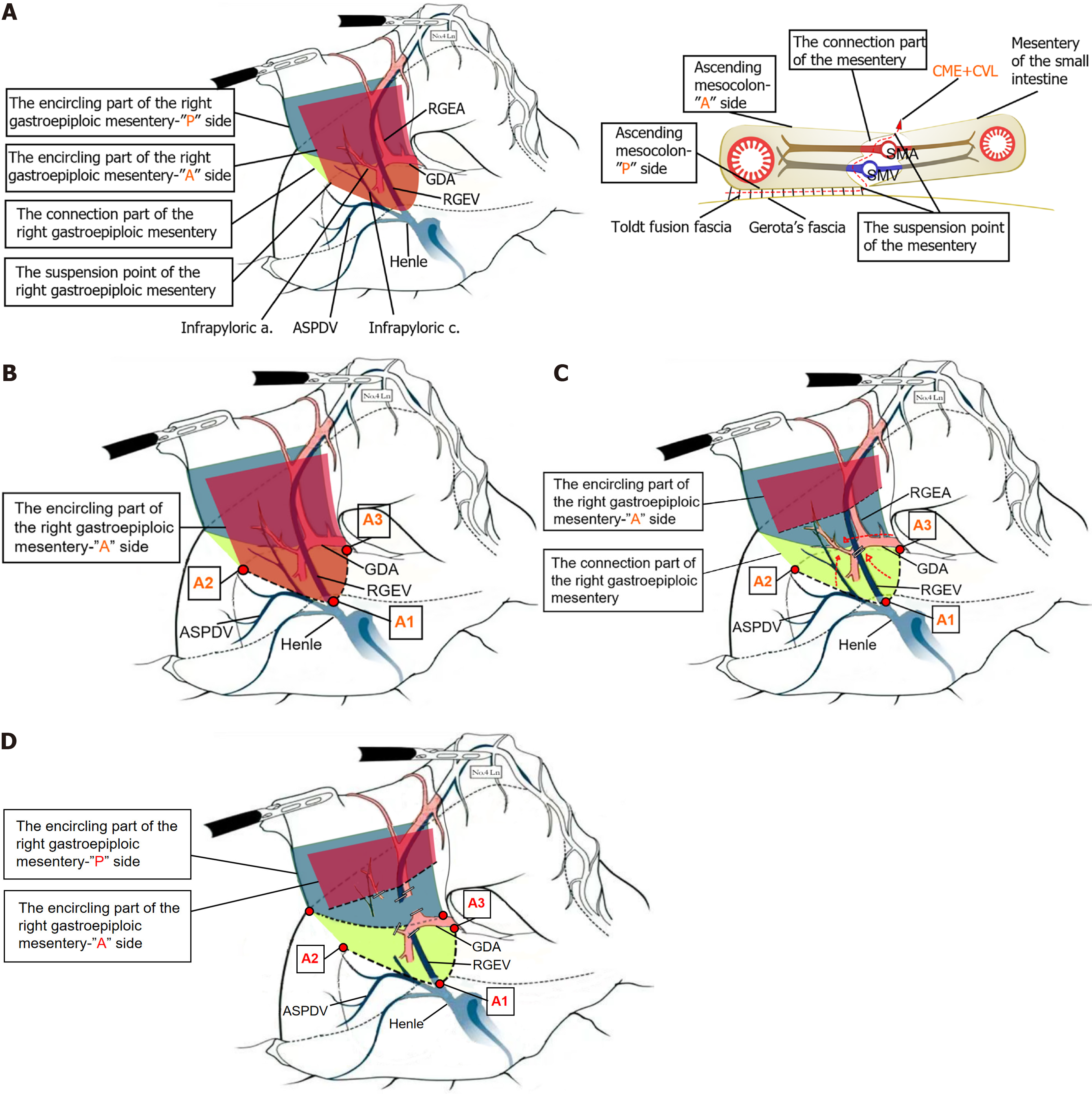
Figure 1 Schematic illustrations of the right gastroepiploic mesentery: Anatomical components, separation, and dissection.
A: Schematic diagram of the three key anatomical components of the right gastroepiploic mesentery; B: Schematic diagram showing the complete separation of the right gastroepiploic mesentery from the transverse mesocolon and identification of the three suspension points of the right gastroepiploic mesentery “anterior” surface; C: Schematic diagram showing the exposure and division of the connection of the right gastroepiploic mesentery; D: Schematic diagram showing the exposure and division of the right gastroepiploic mesentery “posterior” surface. ASPDV: Anterior superior pancreaticoduodenal vein; GDA: Gastroduodenal artery; RGEV: Right gastroepiploic vein; RGEA: Right gastroepiploic arteria; CME: Complete mesogastric excision; CVL: Central vascular ligation; A: Anterior; P: Posterior; A1: First suspended point; A2: Second suspended point; A3: Third suspended point.
- Citation: Pan GF, Zhang WH, Cai ZM, Chen J, Wu JH, Weng JB, Zhu ZP, Guo ZX, Lin JJ, Li ZX, Xu YC. Mesenteric-guided approach to pyloric lymphadenectomy in laparoscopic radical gastrectomy. World J Gastrointest Surg 2025; 17(9): 110064
- URL: https://www.wjgnet.com/1948-9366/full/v17/i9/110064.htm
- DOI: https://dx.doi.org/10.4240/wjgs.v17.i9.110064