
















©The Author(s) 2025.
World J Gastrointest Surg. Sep 27, 2025; 17(9): 109952
Published online Sep 27, 2025. doi: 10.4240/wjgs.v17.i9.109952
Published online Sep 27, 2025. doi: 10.4240/wjgs.v17.i9.109952
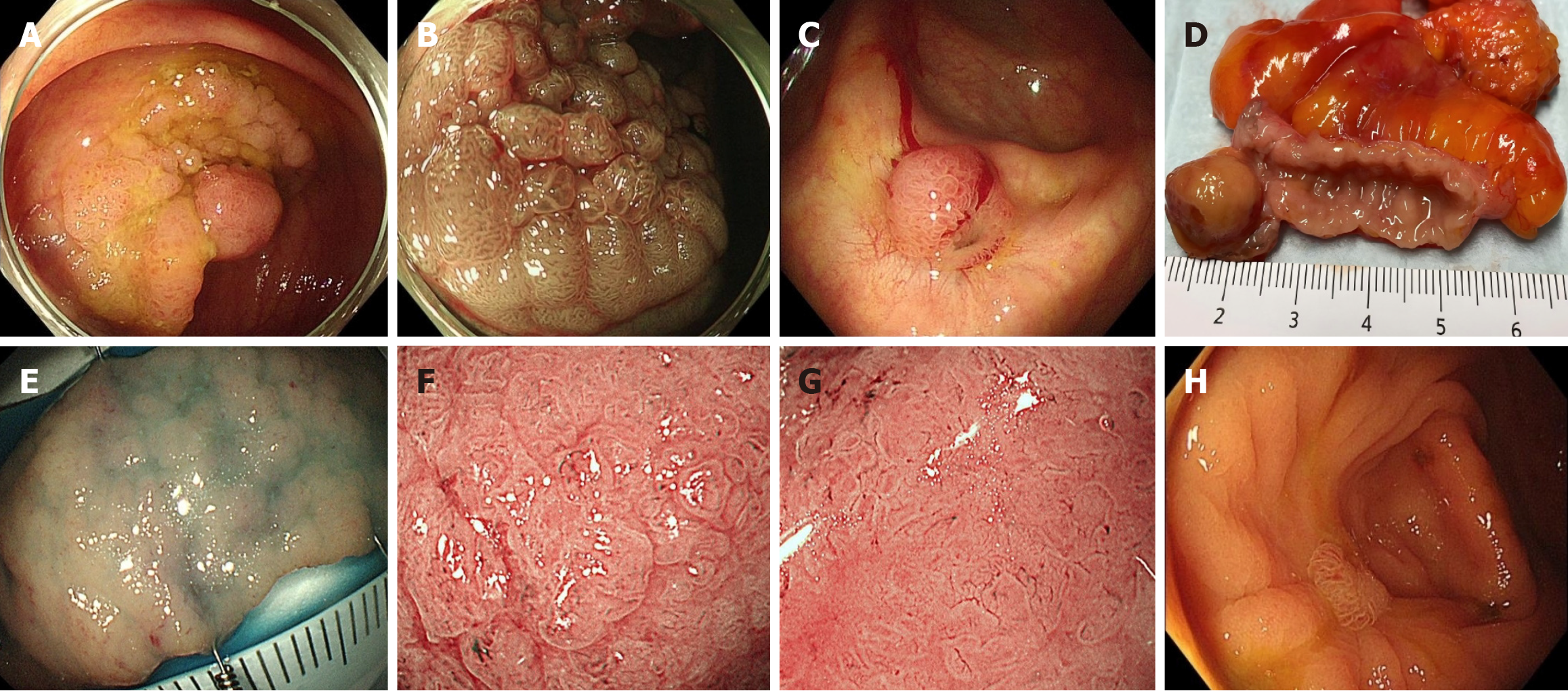
Figure 1 Imaging manifestations.
A: Endoscopic images of the lesion before endoscopic submucosal dissection (ESD); B: Magnifying endoscopy with narrow-band imaging images of the lesion before ESD; C: Endoscopic images after ESD; D: Photographs of longitudinal sections of the gross appendix specimen; E: Endoscopic images of longitudinal sections of the gross appendix specimen; F and G: Magnifying endoscopy with narrow-band imaging images of longitudinal sections of the gross appendix specimen; H: Endoscopic images 1 year after laparoscopic appendectomy showed no signs of recurrence.
- Citation: Huang YH, Ma L, Cao B, Zhang YJ, Gao Q, Zhu ZM, Qiao XL, Wang L, He BG. Endoscopic and laparoscopic treatment of ileocecal laterally spreading tumor with concomitant appendiceal adenoma: A case report and review of literature. World J Gastrointest Surg 2025; 17(9): 109952
- URL: https://www.wjgnet.com/1948-9366/full/v17/i9/109952.htm
- DOI: https://dx.doi.org/10.4240/wjgs.v17.i9.109952