
















©The Author(s) 2025.
World J Gastrointest Surg. Sep 27, 2025; 17(9): 109320
Published online Sep 27, 2025. doi: 10.4240/wjgs.v17.i9.109320
Published online Sep 27, 2025. doi: 10.4240/wjgs.v17.i9.109320
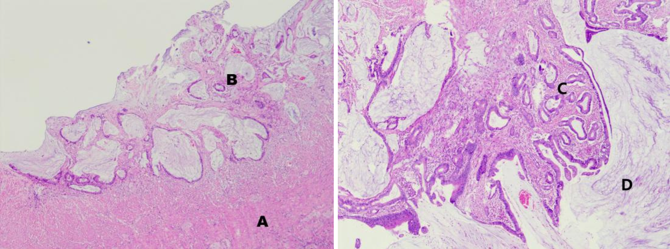
Figure 4 Histological morphology of the appendiceal lesion (hematoxylin-eosin staining).
A: The muscularis propria; B: The submucosal layer, with cancer tissue infiltrating the submucosal layer and muscularis propria, and a significant fibrotic response around the tumor; C: Disorganized structure of cancerous glands, with prominent cellular atypia and abundant mucus secretion; D: Formation of mucin lakes.
- Citation: Guo Q, Lu HY, Lyu H, Tian H, Zhao Q, Zheng YC. Complete appendiceal intussusception and appendiceal mucinous tumor: A case report and review of literature. World J Gastrointest Surg 2025; 17(9): 109320
- URL: https://www.wjgnet.com/1948-9366/full/v17/i9/109320.htm
- DOI: https://dx.doi.org/10.4240/wjgs.v17.i9.109320