
















©The Author(s) 2025.
World J Gastrointest Surg. Sep 27, 2025; 17(9): 109320
Published online Sep 27, 2025. doi: 10.4240/wjgs.v17.i9.109320
Published online Sep 27, 2025. doi: 10.4240/wjgs.v17.i9.109320
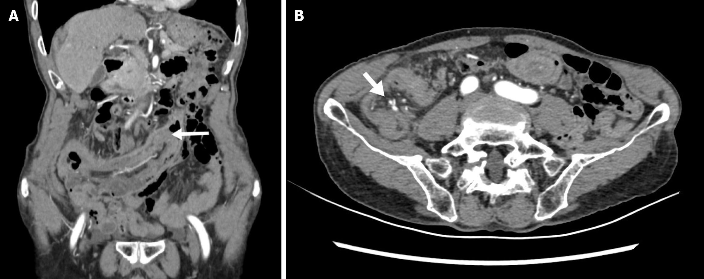
Figure 2 Enhanced abdominal computed tomography.
A: It shows enhanced abdominal computed tomography (CT) in the coronal plane; the white arrow indicates the intussuscepted distal appendix reaching the transverse colon; B: It shows enhanced abdominal CT in the axial plane, the white arrow indicates the ideal blood vessels.
- Citation: Guo Q, Lu HY, Lyu H, Tian H, Zhao Q, Zheng YC. Complete appendiceal intussusception and appendiceal mucinous tumor: A case report and review of literature. World J Gastrointest Surg 2025; 17(9): 109320
- URL: https://www.wjgnet.com/1948-9366/full/v17/i9/109320.htm
- DOI: https://dx.doi.org/10.4240/wjgs.v17.i9.109320