
















©The Author(s) 2025.
World J Gastrointest Surg. Sep 27, 2025; 17(9): 108348
Published online Sep 27, 2025. doi: 10.4240/wjgs.v17.i9.108348
Published online Sep 27, 2025. doi: 10.4240/wjgs.v17.i9.108348
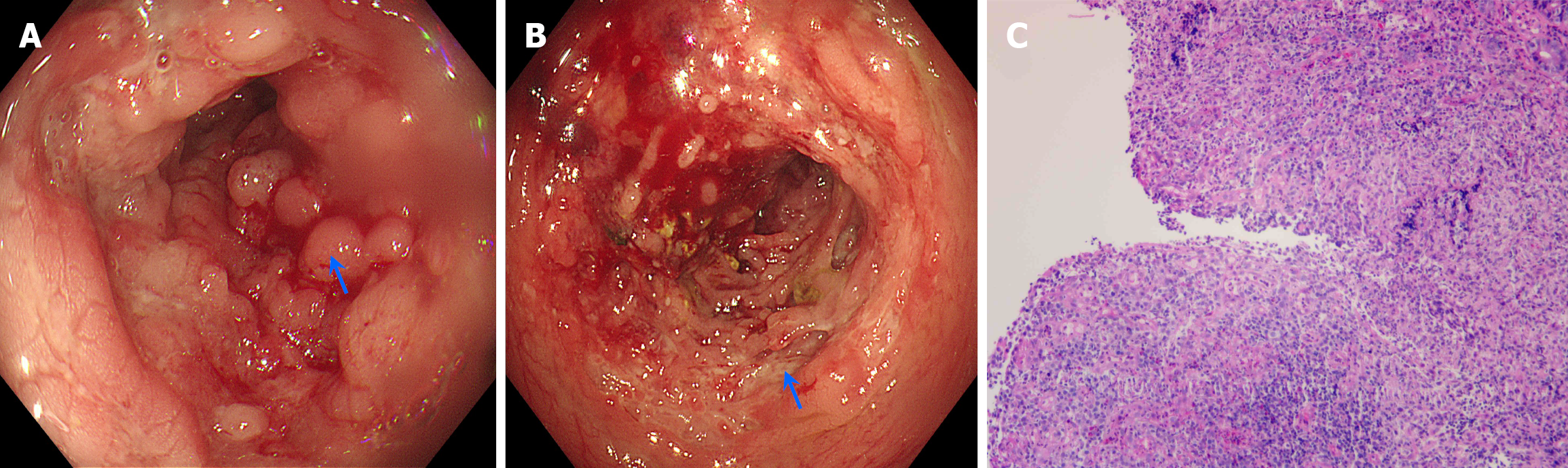
Figure 3 Endoscopic images and intestinal mucosa biopsy histopathology images.
A: Edematous and irregularly hyperplastic intestinal mucosa with a cobblestone appearance under colonoscopic view (arrow); B: Discontinuous creeping ulcers of the intestinal mucosa under colonoscopic view, with normal-appearing mucosa between the ulcers (arrow); C: Histopathological examination revealed lymphocytic aggregation (× 10 magnification, hematoxylin-eosin staining).
- Citation: Wang WQ, Yang JP, Dong JW, Chen YB. Misdiagnosis of Crohn’s disease as appendicitis: A case report. World J Gastrointest Surg 2025; 17(9): 108348
- URL: https://www.wjgnet.com/1948-9366/full/v17/i9/108348.htm
- DOI: https://dx.doi.org/10.4240/wjgs.v17.i9.108348