
















©The Author(s) 2025.
World J Gastrointest Surg. Sep 27, 2025; 17(9): 108348
Published online Sep 27, 2025. doi: 10.4240/wjgs.v17.i9.108348
Published online Sep 27, 2025. doi: 10.4240/wjgs.v17.i9.108348
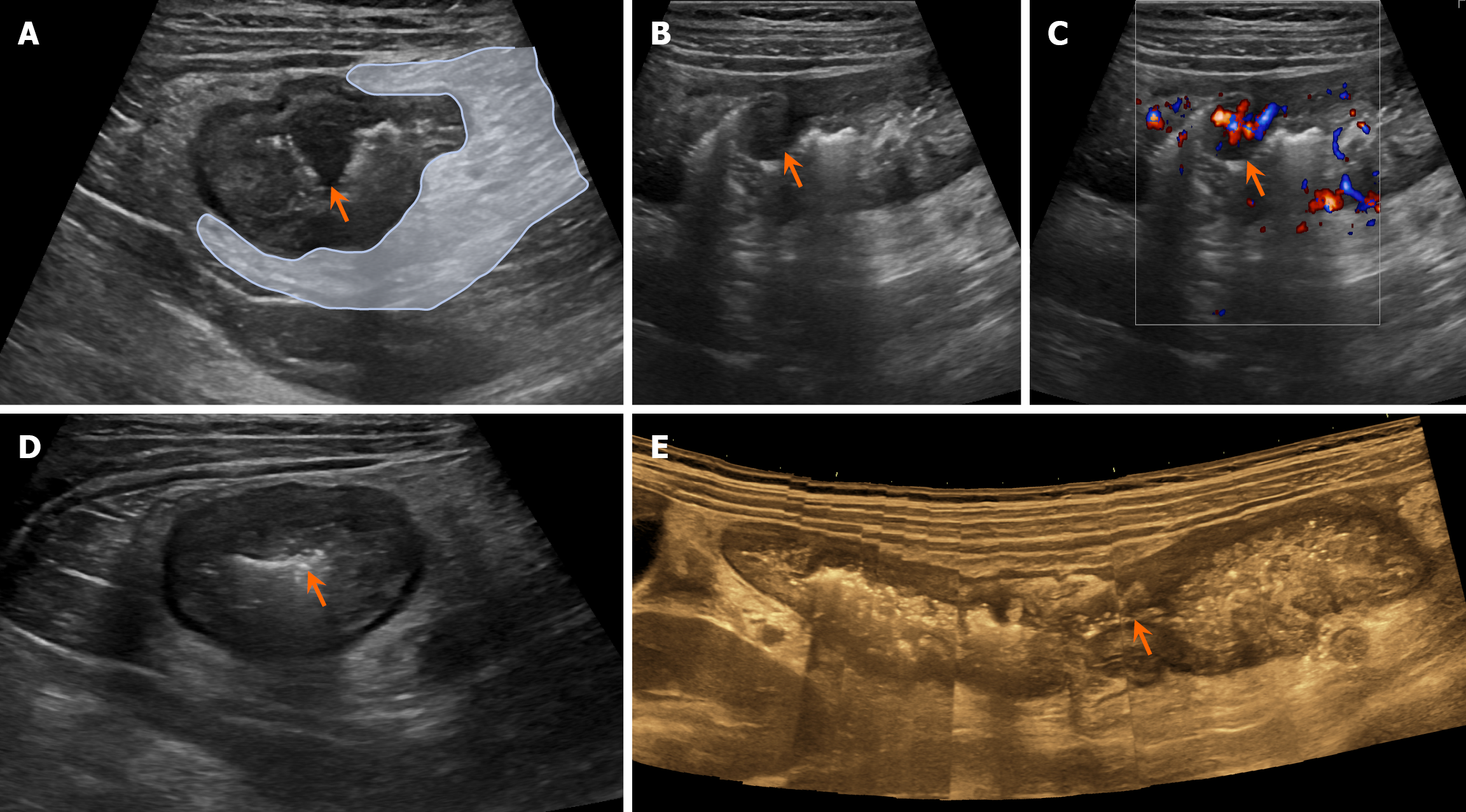
Figure 2 Gastrointestinal ultrasound imaging examinations.
A-C: Inflammatory polyps of the colon with abundant blood flow signals (arrows), and edematous and thickened mesentery (light blue areas); D and E: Luminal stenosis caused by thickening of the ascending colon wall (arrows); Cross-sectional view of the bowel (D); Wide-scene imaging (E).
- Citation: Wang WQ, Yang JP, Dong JW, Chen YB. Misdiagnosis of Crohn’s disease as appendicitis: A case report. World J Gastrointest Surg 2025; 17(9): 108348
- URL: https://www.wjgnet.com/1948-9366/full/v17/i9/108348.htm
- DOI: https://dx.doi.org/10.4240/wjgs.v17.i9.108348