
















©The Author(s) 2025.
World J Gastrointest Surg. Sep 27, 2025; 17(9): 108348
Published online Sep 27, 2025. doi: 10.4240/wjgs.v17.i9.108348
Published online Sep 27, 2025. doi: 10.4240/wjgs.v17.i9.108348
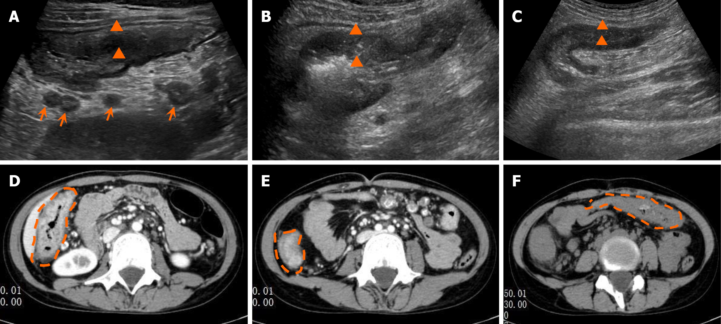
Figure 1 Imaging examinations.
A-C: Gastrointestinal ultrasound imaging showed uneven edema and thickening of the ascending colon, transverse colon, and splenic flexure colon walls (triangle), along with punctate calcifications within the mesenteric lymph nodes due to long-term inflammatory infiltration (arrows); D-F: Computed tomography imaging showed edema and thickening of the ascending colon, ileocecal region, and splenic flexure colon walls (orange dashed lines).
- Citation: Wang WQ, Yang JP, Dong JW, Chen YB. Misdiagnosis of Crohn’s disease as appendicitis: A case report. World J Gastrointest Surg 2025; 17(9): 108348
- URL: https://www.wjgnet.com/1948-9366/full/v17/i9/108348.htm
- DOI: https://dx.doi.org/10.4240/wjgs.v17.i9.108348