
















©The Author(s) 2025.
World J Gastrointest Surg. Sep 27, 2025; 17(9): 107301
Published online Sep 27, 2025. doi: 10.4240/wjgs.v17.i9.107301
Published online Sep 27, 2025. doi: 10.4240/wjgs.v17.i9.107301
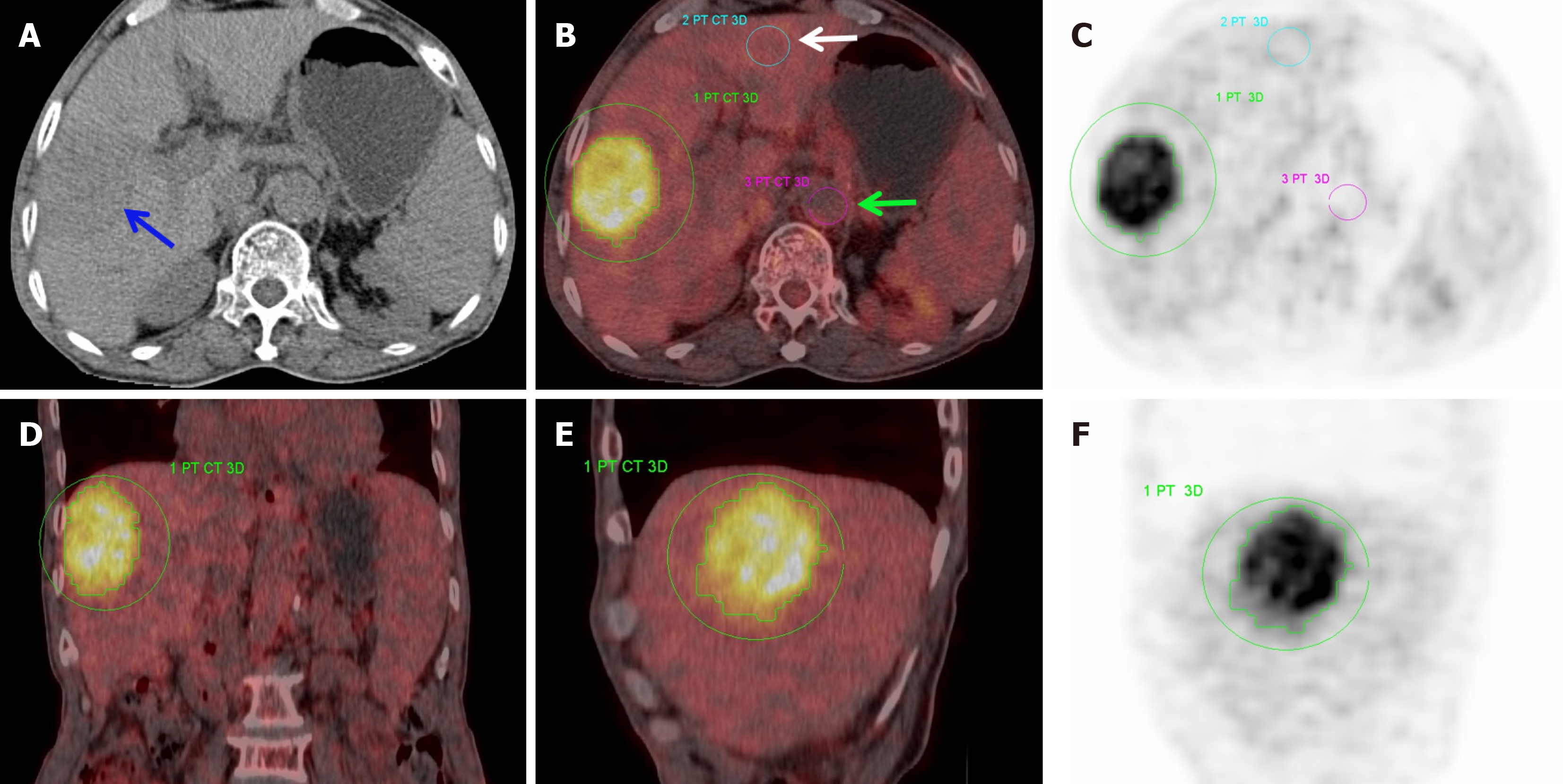
Figure 1 Hepatocellular carcinoma in the right hepatic lobe, with an Edmondson grade of III (Case 1).
The percentage of Ki67 expression was 30%, maximum standardized uptake value was 15.56, mean standardized uptake value was 6.24, metabolic tumor volume was 94.7, total lesion glycolysis was 590.5, tumor-to-normal background ratio was 5.02, and tumor-to-blood pool ratio was 8.64. A: Non-enhanced computerized tomography image depicted a slightly hypodense mass (blue arrow) beneath the capsule of the right hepatic lobe, with an approximate computerized tomography value of 32 Hounsfield unit, a three-dimensional volume of interest was manually outlined along the tumor periphery; B: Normal liver tissue region of interest (white arrow) and abdominal aorta region of interest (green arrow); C: Axial maximum intensity projection image; D: Coronal view; E: Sagittal view; F: Sagittal maximum intensity projection image. PT: Positron tomography; CT: Computed tomography; 3D: Three-dimensional.
- Citation: Niu XB, Li YP, Chao FF, Mei XL, Han XM, Wang RH. Diagnostic significance of fluorine-18-fluorodeoxyglucose-positron emission tomography/computed tomography in differentiating Edmondson grade II and III hepatocellular carcinoma. World J Gastrointest Surg 2025; 17(9): 107301
- URL: https://www.wjgnet.com/1948-9366/full/v17/i9/107301.htm
- DOI: https://dx.doi.org/10.4240/wjgs.v17.i9.107301