
















©The Author(s) 2025.
World J Gastrointest Surg. Sep 27, 2025; 17(9): 107297
Published online Sep 27, 2025. doi: 10.4240/wjgs.v17.i9.107297
Published online Sep 27, 2025. doi: 10.4240/wjgs.v17.i9.107297
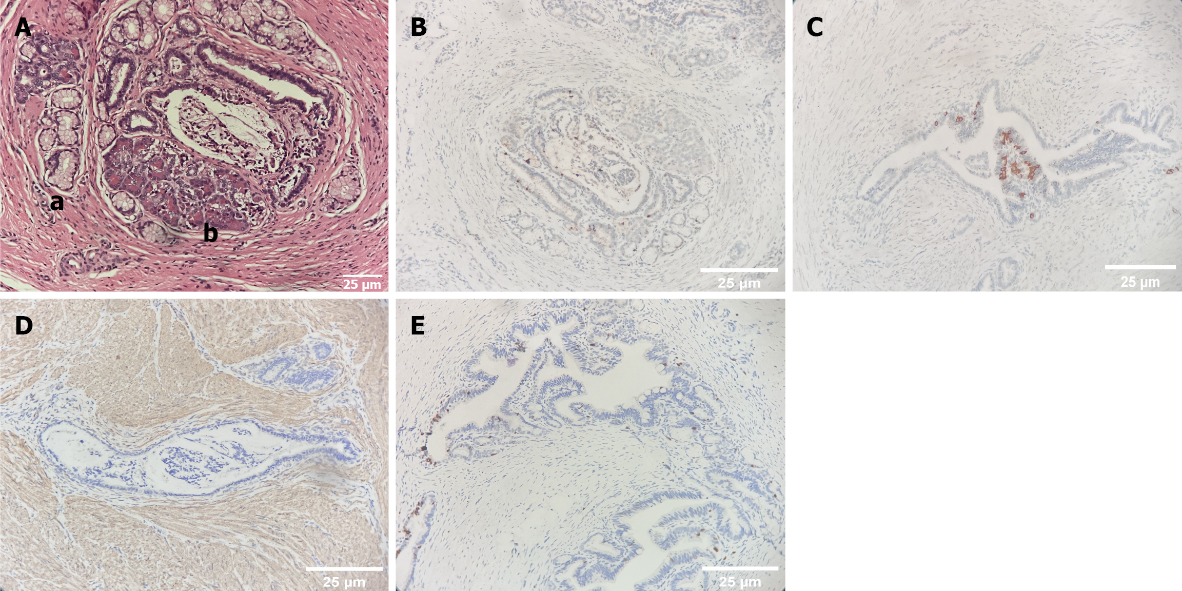
Figure 3 Pathology images for gastric adenomyoma.
A: Hematoxylin and eosin staining, “a” represents intestinal epithelium and “b” indicates ectopic pancreas; B: Representative immunohistochemical staining images of gastrin-positive sections: Gastrin; C: Insulin; D: Smooth muscle actin; E: Ki-67 staining.
- Citation: Zhang J, Sun S, Zheng S, Ma YY, Chen G. Gastric adenomyoma in children: A case report. World J Gastrointest Surg 2025; 17(9): 107297
- URL: https://www.wjgnet.com/1948-9366/full/v17/i9/107297.htm
- DOI: https://dx.doi.org/10.4240/wjgs.v17.i9.107297