
















©The Author(s) 2025.
World J Gastrointest Surg. Sep 27, 2025; 17(9): 107139
Published online Sep 27, 2025. doi: 10.4240/wjgs.v17.i9.107139
Published online Sep 27, 2025. doi: 10.4240/wjgs.v17.i9.107139
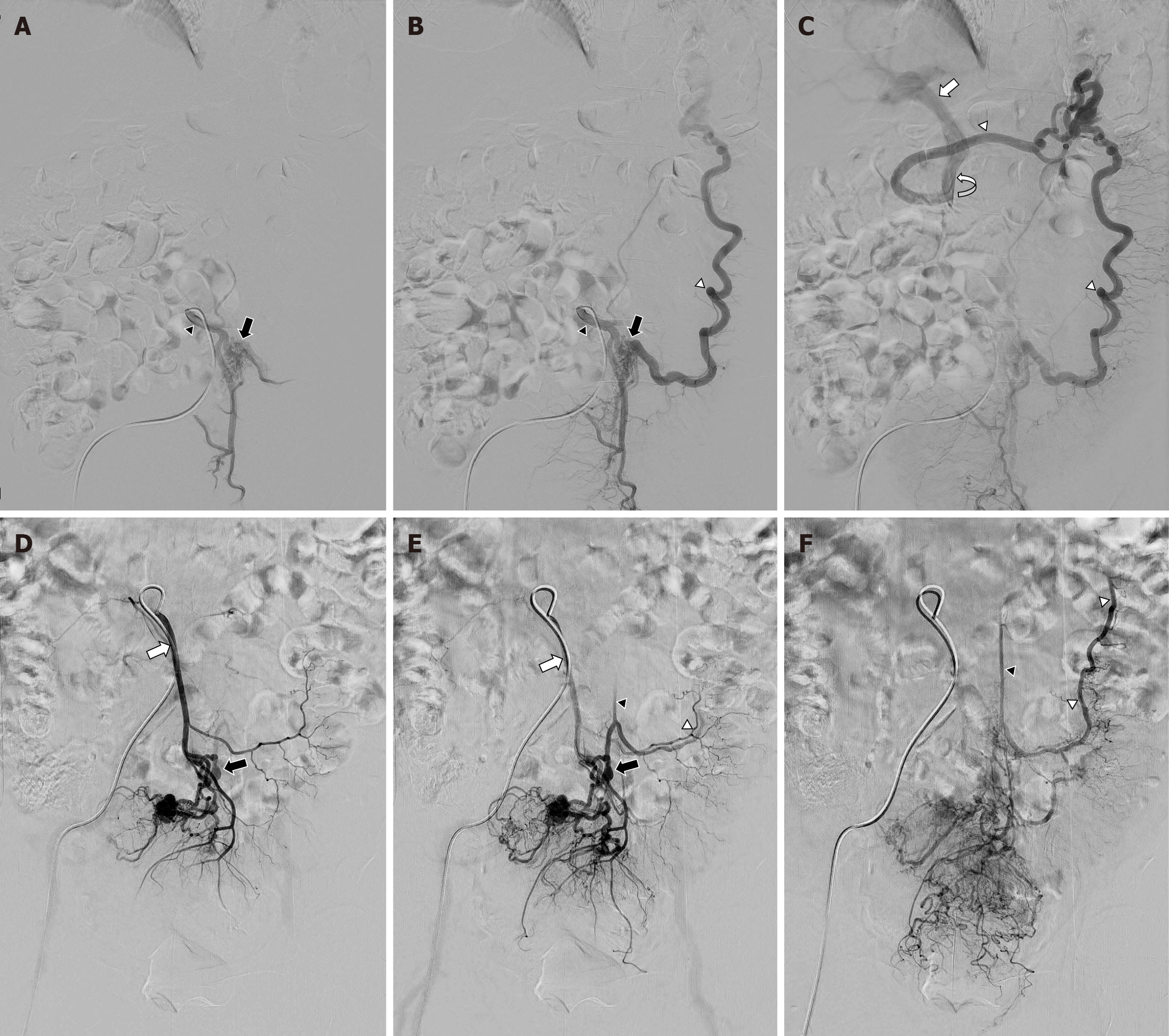
Figure 4 Angiography of the inferior mesenteric artery.
A: Nidus of the fistula (black arrows) supplied by the inferior mesenteric artery (black arrowheads); B: Prompt filling of the dilated marginal vein (white arrowheads) with drainage; C: The superior mesenteric vein (white curved arrow) and portal vein (white arrow). Invisible occluded inferior mesenteric vein; D: Nidus (black arrows) observed in the distal portion of the inferior mesenteric artery (white arrows); E: Early visualization of the inferior mesenteric vein (black arrowheads); F: Marginal vein (white arrowheads), with both showing a beaded appearance.
- Citation: Moon YJ, Lee SH. Inferior mesenteric arteriovenous fistula: Two case reports. World J Gastrointest Surg 2025; 17(9): 107139
- URL: https://www.wjgnet.com/1948-9366/full/v17/i9/107139.htm
- DOI: https://dx.doi.org/10.4240/wjgs.v17.i9.107139