
















©The Author(s) 2025.
World J Gastrointest Surg. Sep 27, 2025; 17(9): 107139
Published online Sep 27, 2025. doi: 10.4240/wjgs.v17.i9.107139
Published online Sep 27, 2025. doi: 10.4240/wjgs.v17.i9.107139
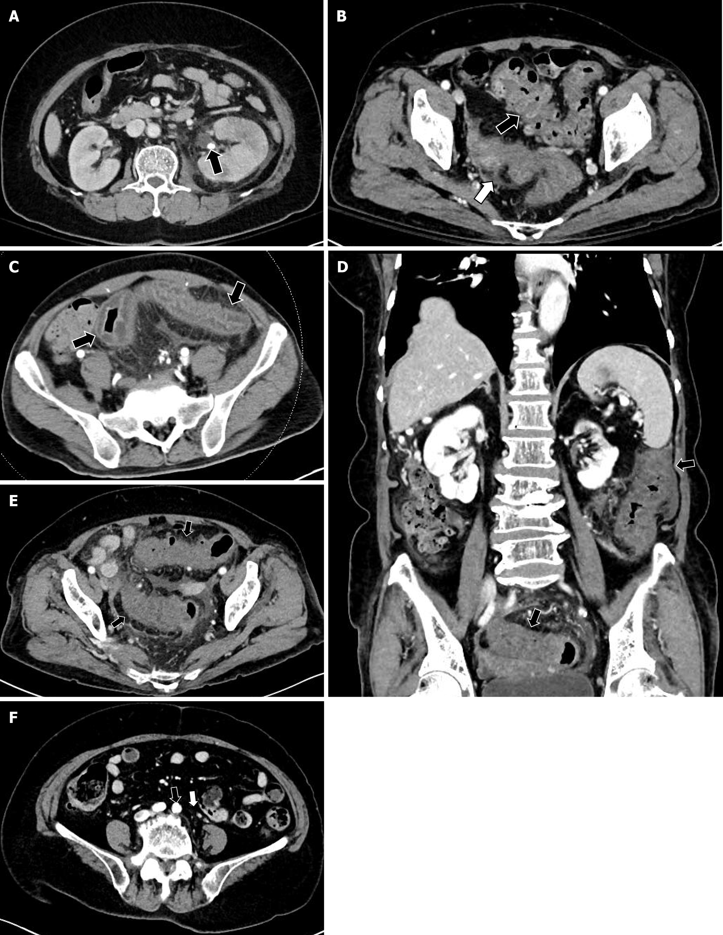
Figure 1 Multi-detector computed tomography scan.
A: Multifocal low densities in the enlarged left kidney with ureteral dilatation, periureteric and perirenal fat stranding, and a suspected pelvic stone (arrow), indicative of acute pyelonephritis; B: Sigmoid colon diverticulitis with wall thickening (black arrow), pericolic fat stranding, and mild fluid collection (white arrow); C: Edematous, thickened, and hypoenhanced wall of the sigmoid colon with adjacent fat stranding (arrows) observed in the arterial phase; D: Long-segment colonic wall thickening with reduced enhancement, submucosal edema, pericolic fat stranding; E: Mild fluid collection extending from the splenic flexure to the upper rectum (black arrows); F: No communication observed between the inferior mesenteric artery (black arrow) and vein (white arrow).
- Citation: Moon YJ, Lee SH. Inferior mesenteric arteriovenous fistula: Two case reports. World J Gastrointest Surg 2025; 17(9): 107139
- URL: https://www.wjgnet.com/1948-9366/full/v17/i9/107139.htm
- DOI: https://dx.doi.org/10.4240/wjgs.v17.i9.107139