Copyright
©The Author(s) 2025.
World J Gastrointest Surg. Jul 27, 2025; 17(7): 107804
Published online Jul 27, 2025. doi: 10.4240/wjgs.v17.i7.107804
Published online Jul 27, 2025. doi: 10.4240/wjgs.v17.i7.107804
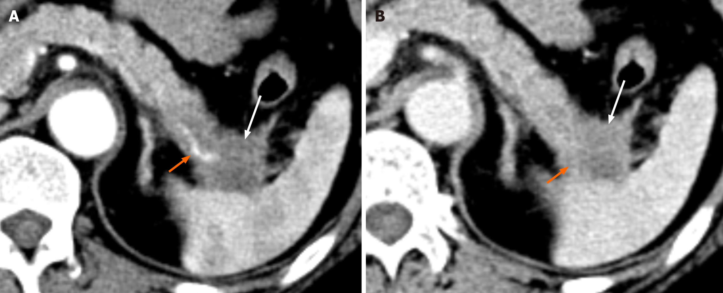
Figure 3 Computed tomography images of a 61-year-old female with resectable pancreatic ductal adenocarcinoma.
A: Arterial phase; B: Portal venous phase axial contrast-enhanced computed tomography images revealed a 3.8 cm hypodense mass in the tail of the pancreas (indicated by a white arrow) with invasion of the splenic artery (indicated by a orange arrow). The image scale was approximately 0.31 mm per pixel. There was no evidence of tumor necrosis or suspicious metastatic lymph nodes. With a risk score of 3, the patient was placed in the moderate prognosis group. A standard distal pancreatectomy was performed, and the tumor recurred 18 months postoperatively.
- Citation: Liu XH, Xie JH, Zhu XS, Liu LH. Preoperative computed tomography-based risk stratification model validation for postoperative pancreatic ductal adenocarcinoma recurrence. World J Gastrointest Surg 2025; 17(7): 107804
- URL: https://www.wjgnet.com/1948-9366/full/v17/i7/107804.htm
- DOI: https://dx.doi.org/10.4240/wjgs.v17.i7.107804


