Copyright
©The Author(s) 2025.
World J Gastrointest Surg. Jul 27, 2025; 17(7): 105956
Published online Jul 27, 2025. doi: 10.4240/wjgs.v17.i7.105956
Published online Jul 27, 2025. doi: 10.4240/wjgs.v17.i7.105956
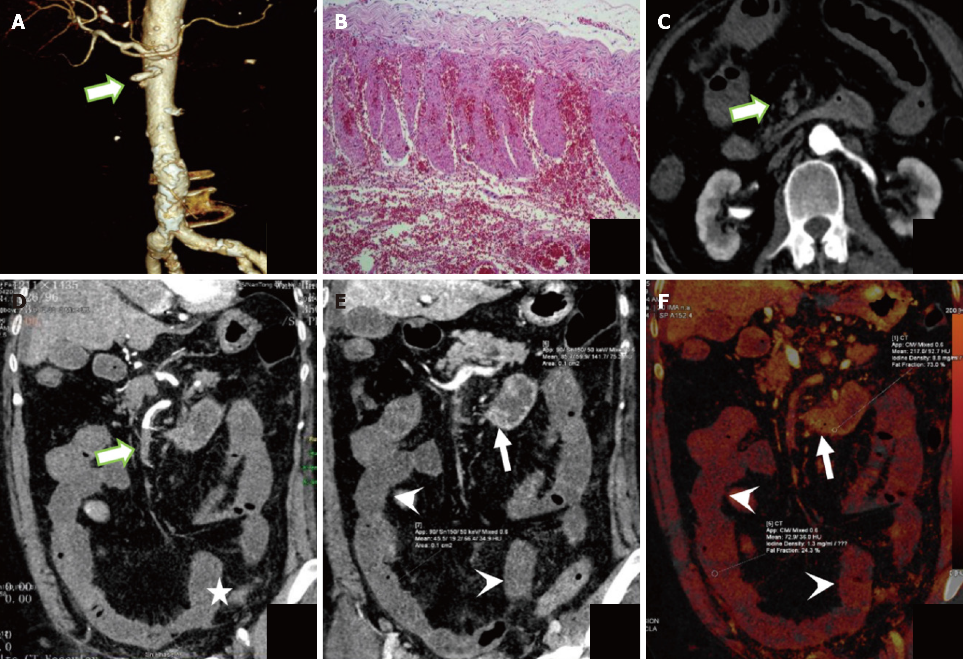
Figure 7 The representative dual energy computed tomography image of a 75-year-old woman with irreversible transmural intestinal necrosis.
A: Volume rendering technique image; B: Hematoxylin and eosin staining images; C and D: Transverse and coronal images of the portal vein phase at 120 kVp; E: Portal vein phase at 50 keV image; F: Iodine density. Dual energy computed tomography examination reveals filling defect in superior mesenteric artery (area I+ II + III + IV) (thick arrow in A, C, and D), decreased and absent diffuse intestinal wall enhancement (arrowhead in E and F), normal intestinal duct (thin arrow in E and F), intestinal dilatation (star in D). Computed tomography 50 keV normal/lesion = 2.13, iodine concentration normal/lesion = 6.77.
- Citation: Yang JS, Xu ZY, Chen FX, Wang MR, Fan XL, He BS. Diagnostic value of dual-energy computed tomography in irreversible transmural intestinal necrosis in patients with acute occlusive mesenteric ischemia. World J Gastrointest Surg 2025; 17(7): 105956
- URL: https://www.wjgnet.com/1948-9366/full/v17/i7/105956.htm
- DOI: https://dx.doi.org/10.4240/wjgs.v17.i7.105956


