Copyright
©The Author(s) 2025.
World J Gastrointest Surg. Jul 27, 2025; 17(7): 105956
Published online Jul 27, 2025. doi: 10.4240/wjgs.v17.i7.105956
Published online Jul 27, 2025. doi: 10.4240/wjgs.v17.i7.105956
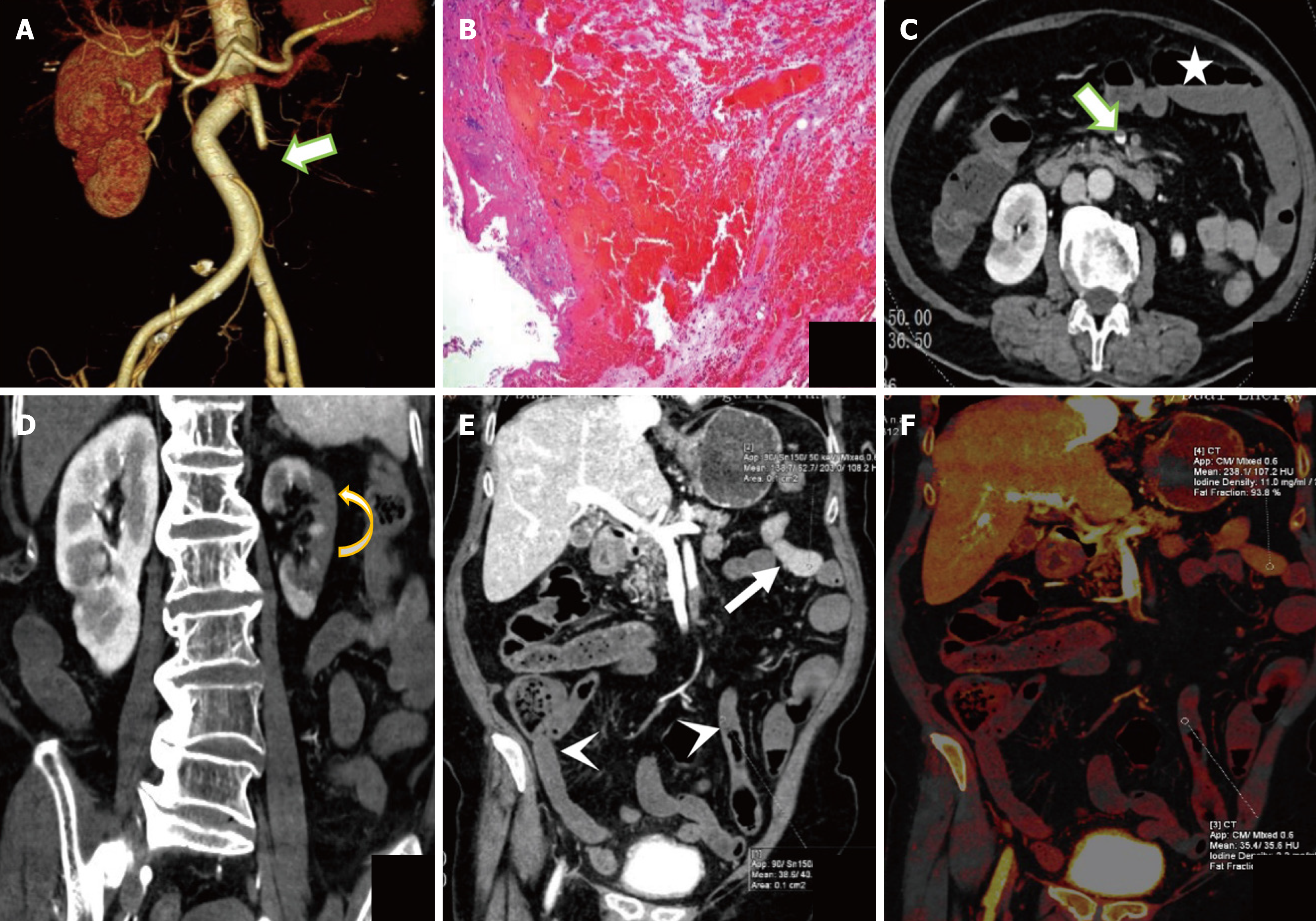
Figure 6 The representative dual energy computed tomography image of a 76-year-old woman with irreversible transmural intestinal necrosis.
A: Volume rendering technique image; B: Hematoxylin and eosin staining images; C and D: Transverse and coronal images of the portal vein phase at 120 kVp; E: Portal vein phase at 50 keV image; F: Iodine density. Dual energy computed tomography examination reveals filling defect in superior mesenteric artery (area II + III + IV) and superior mesenteric vein (thick arrow in A and C), decreased and absent diffuse intestinal wall enhancement (arrowhead in E), normal intestinal duct (thin arrow in E), intestinal dilatation (star in C), and left renal infarction (curved arrow in D). Computed tomography 50 keV normal/lesion = 4.33, iodine concentration normal/lesion = 5.00.
- Citation: Yang JS, Xu ZY, Chen FX, Wang MR, Fan XL, He BS. Diagnostic value of dual-energy computed tomography in irreversible transmural intestinal necrosis in patients with acute occlusive mesenteric ischemia. World J Gastrointest Surg 2025; 17(7): 105956
- URL: https://www.wjgnet.com/1948-9366/full/v17/i7/105956.htm
- DOI: https://dx.doi.org/10.4240/wjgs.v17.i7.105956


